2,859
Views
15
CrossRef citations to date
0
Altmetric
Report

Evaluation of a glycoengineered monoclonal antibody via LC-MS analysis in combination with multiple enzymatic digestion

, , , , &
Pages 340-346 | Received 25 Sep 2015, Accepted 21 Oct 2015, Published online: 09 Dec 2015

Figures & data

Figure 1. Glycosylation remodeling of rituximab to prepare rituximab with homogenous N-glycan with 2,6 linked sialic acid using EndoS or EndoS2.

Figure 1. Glycosylation remodeling of rituximab to prepare rituximab with homogenous N-glycan with 2,6 linked sialic acid using EndoS or EndoS2.

Table 1. Summary of rituximab N-glycan.

Figure 2. HPLC-FLD profile of procainamide labeled N-glycans from rituximab and the glycoengineered rituximab with α2,6 linked sialic acid. A). HPLC profile of rituximab N-glycans with major species shown. B). The glycan profile from the glycoengineered rituximab prepared by Endo S digestion. The major glycan is G2FS2 with α2,6 linked sialic acid. C). The glycan profile from the glycoengineered rituximab after α2-3 Neuraminidase S treatment. D). The glycan profile from the glycoengineered rituximab after α2-3,6,8,9 Neuraminidase A treatment. E). The glycan profile from the glycoengineered rituximab prepared by Endo S2 digestion.
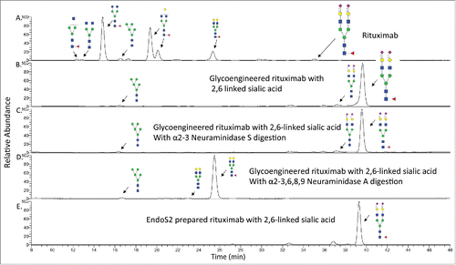
Figure 2. HPLC-FLD profile of procainamide labeled N-glycans from rituximab and the glycoengineered rituximab with α2,6 linked sialic acid. A). HPLC profile of rituximab N-glycans with major species shown. B). The glycan profile from the glycoengineered rituximab prepared by Endo S digestion. The major glycan is G2FS2 with α2,6 linked sialic acid. C). The glycan profile from the glycoengineered rituximab after α2-3 Neuraminidase S treatment. D). The glycan profile from the glycoengineered rituximab after α2-3,6,8,9 Neuraminidase A treatment. E). The glycan profile from the glycoengineered rituximab prepared by Endo S2 digestion.

Figure 3. The detection of pyro-E in rituximab. A). Precursor mass of pyro-glutamic acid of the heavy chain from rituximab. B). CID-MSCitation2 of the precursor mass from . The theoretical and observed monoisotopic mass are indicated in the figure.
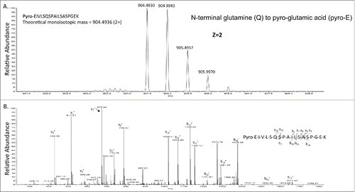
Figure 3. The detection of pyro-E in rituximab. A). Precursor mass of pyro-glutamic acid of the heavy chain from rituximab. B). CID-MSCitation2 of the precursor mass from Figure 3A. The theoretical and observed monoisotopic mass are indicated in the figure.
Supplemental material

Liu et al Supplemental Data

Download Zip (609.8 KB)