1,991
Views
22
CrossRef citations to date
0
Altmetric
Reports

A bispecific antibody effectively neutralizes all four serotypes of dengue virus by simultaneous blocking virus attachment and fusion

, , , , , , , , , , , , , , , , & show all
Pages 574-584 | Received 08 Sep 2015, Accepted 25 Jan 2016, Published online: 10 Mar 2016

Figures & data

Figure 1. Structure and characterization of DVD-Ig. (A) Schematic representation of the structure of DVD-1A1D-2A10. (B) SDS-PAGE analysis of purified antibodies under non-reducing and reducing conditions. Lane 1, molecular weight protein markers; lane 2, DVD-1A1D-2A10; lane 3, bivalent antibody.
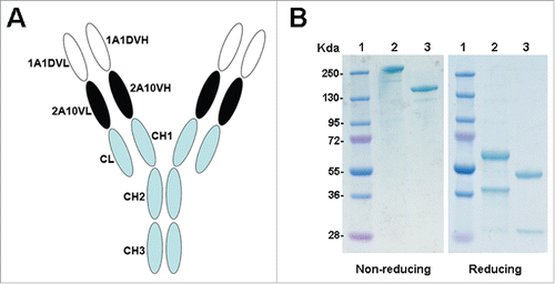
Figure 1. Structure and characterization of DVD-Ig. (A) Schematic representation of the structure of DVD-1A1D-2A10. (B) SDS-PAGE analysis of purified antibodies under non-reducing and reducing conditions. Lane 1, molecular weight protein markers; lane 2, DVD-1A1D-2A10; lane 3, bivalent antibody.

Figure 2. Bispecific binding capacity of DVD-1A1D-2A10. (A) Western blot analysis of DVD-1A1D-2A10. 1A1D and 2A10 bind DIII and DI/II, respectively; DVD-Ig binds both D I/II and D III. (B) A competition ELISA was used to confirm DVD-1A1D-2A10 binding D I/II and D III simultaneously. (C) Bispecific binding to virus. BHK21 cells were infected with DENV2-43. Three to 5 days after infection, cells were fixed and analyzed by indirect immunofluorescence analysis. First, cells were blocked by murine antibodies (m1A1D, m2A10 and m1A1D+m2A10, respectively). Then, these cells were incubated with corresponding chimeric antibodies (c1A1D, c2A10 and cDVD-1A1D-2A10) and tested by fluorescein isothiocyanate (FITC)–conjugated anti-human IgG. Green means that chimeric antibody could bind to E proteins on the virus surface. Red represent negative cells. (D and E). Binding activity of DVD-1A1D-2A10 compared to parental antibodies. Increasing concentrations of DVD-1A1D-2A10, 1A1D, and 2A10 were added to 96-well plates coated with DI/II (D) or DIII (E). DVD-1A1D-2A10 retained equal binding activity of 1A1D and 2A10, respectively.
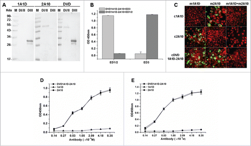
Figure 2. Bispecific binding capacity of DVD-1A1D-2A10. (A) Western blot analysis of DVD-1A1D-2A10. 1A1D and 2A10 bind DIII and DI/II, respectively; DVD-Ig binds both D I/II and D III. (B) A competition ELISA was used to confirm DVD-1A1D-2A10 binding D I/II and D III simultaneously. (C) Bispecific binding to virus. BHK21 cells were infected with DENV2-43. Three to 5 days after infection, cells were fixed and analyzed by indirect immunofluorescence analysis. First, cells were blocked by murine antibodies (m1A1D, m2A10 and m1A1D+m2A10, respectively). Then, these cells were incubated with corresponding chimeric antibodies (c1A1D, c2A10 and cDVD-1A1D-2A10) and tested by fluorescein isothiocyanate (FITC)–conjugated anti-human IgG. Green means that chimeric antibody could bind to E proteins on the virus surface. Red represent negative cells. (D and E). Binding activity of DVD-1A1D-2A10 compared to parental antibodies. Increasing concentrations of DVD-1A1D-2A10, 1A1D, and 2A10 were added to 96-well plates coated with DI/II (D) or DIII (E). DVD-1A1D-2A10 retained equal binding activity of 1A1D and 2A10, respectively.

Figure 3. Enhanced neutralizing ability of DVD in vitro. DENV1-4 viruses were incubated with antibodies at different concentrations. Neutralization abilities were evaluated by PRNT assays using BHK21 cells. Results were exhibited by percentage of plaques reduction and shown as mean ± SD of 3 independent experiments.
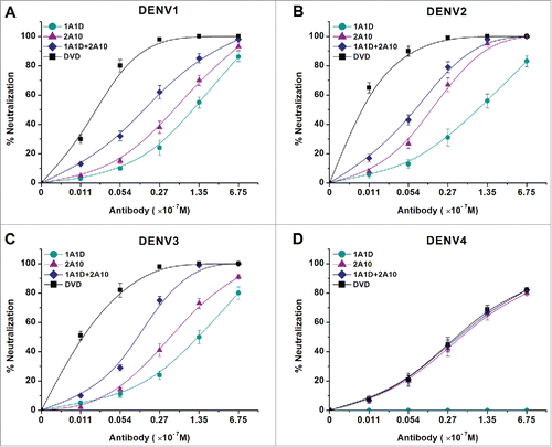
Figure 3. Enhanced neutralizing ability of DVD in vitro. DENV1-4 viruses were incubated with antibodies at different concentrations. Neutralization abilities were evaluated by PRNT assays using BHK21 cells. Results were exhibited by percentage of plaques reduction and shown as mean ± SD of 3 independent experiments.

Table 1. PRNT50 values of antibodies against DENV1-4.

Figure 4. DVD-1A1D-2A10 could neutralize DENV infection by blocking both viral adsorption and fusion. Regardless of blocking absorption (A) or fusion (B), DVD-1A1D-2A10 was more efficient than antibodies mixture or single mAb. In contrast, simply mixing 2 mAbs was not significantly better than single mAb. Neutralization percentages at the different antibody concentrations are shown as mean ± SD of 3 independent experiments.
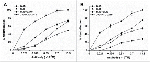
Figure 4. DVD-1A1D-2A10 could neutralize DENV infection by blocking both viral adsorption and fusion. Regardless of blocking absorption (A) or fusion (B), DVD-1A1D-2A10 was more efficient than antibodies mixture or single mAb. In contrast, simply mixing 2 mAbs was not significantly better than single mAb. Neutralization percentages at the different antibody concentrations are shown as mean ± SD of 3 independent experiments.

Figure 5. Elimination of ADE in vitro. Serial antibody dilutions were incubated with DENV1-4 before added to K562 cells. The antibody dilutions were expressed in logarithms. Results were shown by the number of plaques. The data were shown as mean ± SD of 3 independent experiments.
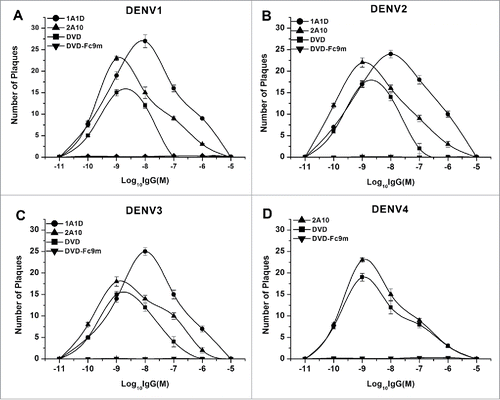
Figure 5. Elimination of ADE in vitro. Serial antibody dilutions were incubated with DENV1-4 before added to K562 cells. The antibody dilutions were expressed in logarithms. Results were shown by the number of plaques. The data were shown as mean ± SD of 3 independent experiments.

Figure 6. Enhanced neutralizing ability of DVD-1A1D-2A10 against DENV1-4 in vivo. Antibodies were diluted to corresponding concentrations, then pre-mixed with different virus. The mixture was inoculated into suckling mice by intracranial injection. Each group contains 8 to 12 suckling mice. Data only showed the final survival rate in percentage of the Kaplan-Meier survival curves, and the negative control mice (not shown) were dead 6 to 8 days after infection. The log-rank test was used to analyze the differences between the DVD groups and other antibodies treated groups. Significant differences are indicated by asterisks (*** P < 0.001, ** P < 0.01 and * P < 0.05).
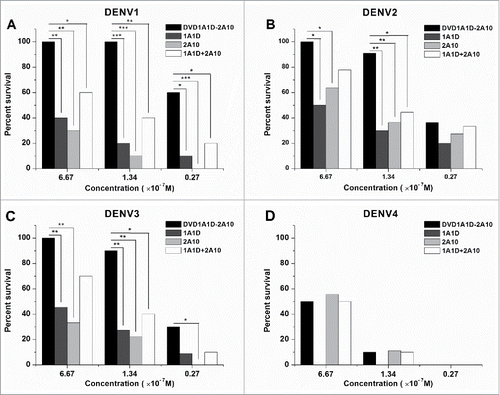
Figure 6. Enhanced neutralizing ability of DVD-1A1D-2A10 against DENV1-4 in vivo. Antibodies were diluted to corresponding concentrations, then pre-mixed with different virus. The mixture was inoculated into suckling mice by intracranial injection. Each group contains 8 to 12 suckling mice. Data only showed the final survival rate in percentage of the Kaplan-Meier survival curves, and the negative control mice (not shown) were dead 6 to 8 days after infection. The log-rank test was used to analyze the differences between the DVD groups and other antibodies treated groups. Significant differences are indicated by asterisks (*** P < 0.001, ** P < 0.01 and * P < 0.05).

Figure 7. Preventive effects in vivo. Groups of one day old suckling mice were administrated with 50 μg of antibodies one day before challenge with 200 PFU of DENV1-4, respectively. PBS replaced antibody in the negative controls. The number of animals for each group ranged from 9 to 12. Kaplan-Meier survival curves were analyzed by the log-rank test and compared to curves of the PBS controls. Significant differences are indicated by asterisks (*** P < 0.001, ** P < 0.01 and * P < 0.05).
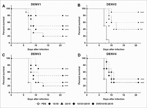
Figure 7. Preventive effects in vivo. Groups of one day old suckling mice were administrated with 50 μg of antibodies one day before challenge with 200 PFU of DENV1-4, respectively. PBS replaced antibody in the negative controls. The number of animals for each group ranged from 9 to 12. Kaplan-Meier survival curves were analyzed by the log-rank test and compared to curves of the PBS controls. Significant differences are indicated by asterisks (*** P < 0.001, ** P < 0.01 and * P < 0.05).
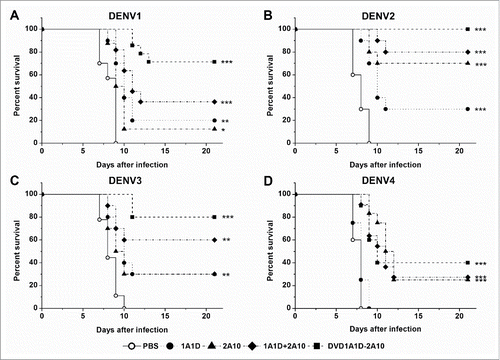
Figure 8. Therapeutic effects in vivo. Groups of one day old suckling mice were administrated with 50 μg of antibodies 4 hours after challenge with 200 PFU of DENV1-4, respectively. PBS replaced antibody in the negative controls. The number of animals for each group ranged from 9 to 12. Kaplan-Meier survival curves were analyzed by the log-rank test and compared to curves of the PBS controls. Significant differences are indicated by asterisks (*** P < 0.001, ** P < 0.01 and * P < 0.05).

Figure 8. Therapeutic effects in vivo. Groups of one day old suckling mice were administrated with 50 μg of antibodies 4 hours after challenge with 200 PFU of DENV1-4, respectively. PBS replaced antibody in the negative controls. The number of animals for each group ranged from 9 to 12. Kaplan-Meier survival curves were analyzed by the log-rank test and compared to curves of the PBS controls. Significant differences are indicated by asterisks (*** P < 0.001, ** P < 0.01 and * P < 0.05).

Reprints and Corporate Permissions

Please note: Selecting permissions does not provide access to the full text of the article, please see our help page How do I view content?

To request a reprint or corporate permissions for this article, please click on the relevant link below:

Academic Permissions

Please note: Selecting permissions does not provide access to the full text of the article, please see our help page How do I view content?

Obtain permissions instantly via Rightslink by clicking on the button below:

If you are unable to obtain permissions via Rightslink, please complete and submit this Permissions form. For more information, please visit our Permissions help page.