Figures & data
Figure 1. Deconvoluted LCMS spectra of reduced, control and oxidized infliximab. (A) Fc; (B) LC; (C) Fd.
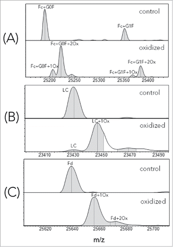
Figure 2. Consolidated forced oxidation experimental results for both Fd and LC.
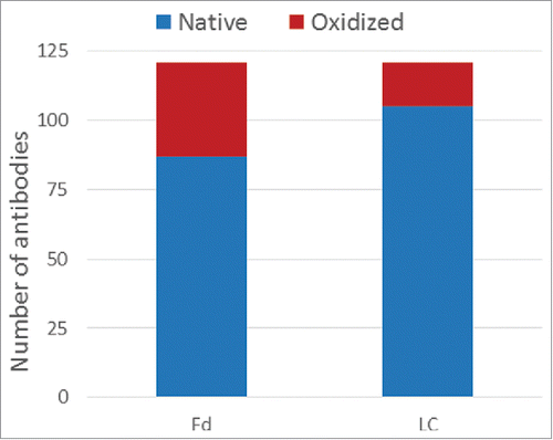
Figure 3. Determination of optimal predicted SASA cutoff.
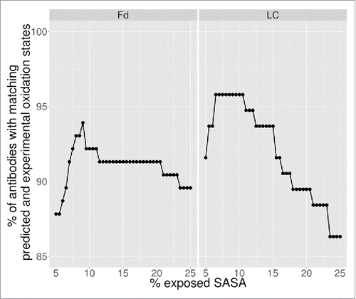
Table 1. Comparison of experimental assignments versus computational predictions at 9% SASA cutoff.
Table 2. List of 11 false negatives in Fd and light chain.
Figure 4. High methionine SASA (> 9%) is indicative of HC (Fd) and LC oxidation event(s).
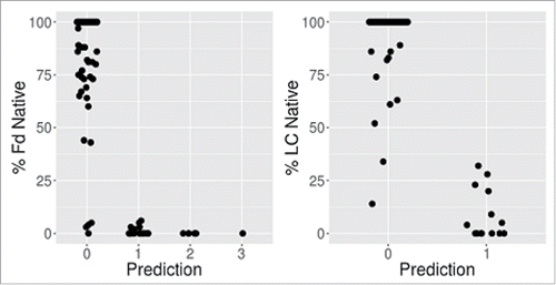
Table 3. Peptide mapping MS2 analysis of selected samples with false negative occurrences.
Table 4. Methionine near the end of H3.
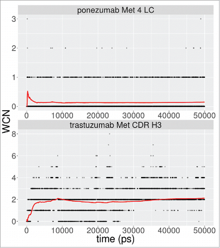
Figure 5. Water coordination number for methionine sulfur atom in a shell of radius 5.5Å. The red lines denote the running average over the simulation.