Figures & data
Figure 1. N-glycan profiles, %Galactosylated, %Afucosylated and %High Mannosylated, and %Charged levels of Remicade®, Flixabi®, and Remsima®. (A) N-glycan profiles of Remicade®, Flixabi®, and Remsima®. EU, emission unit. (B) Percentage of galactosylated glycans. (C) Percentage of afucose and high mannosylated glycans. (D) Percentage of charged glycans. Light color dots: Individual data; Dark color dots: Mean data with mean value and standard deviation; Bar: 95% confidence interval of mean.
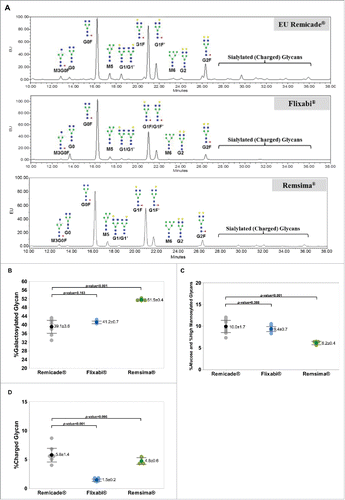
Figure 2. FcγRIIIa binding activity of Remicade®, Flixabi®, and Remsima®. (A) Percentage of relative binding activity using surface plasmon resonance. (B) Percentage of relative binding activity using AlphaScreen. (C) Percentage of relative binding activity using fluorescence-activated cell sorting. Light color dots: Individual data; Dark color dots: Mean data with mean value and standard deviation; Bar: 95% confidence interval of mean.
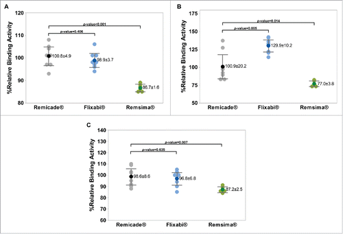
Figure 3. Antibody-dependent cell-mediated cytotoxicity of Remicade®, Flixabi®, and Remsima®. (A) CD16-positive cells (Count %) and mean fluorescence intensities (MFI) in CD3 negative populations from healthy donor peripheral blood mononuclear cells and NK92-CD16a cell line. APC, Allophycocyanin; APC-A, Area of APC detection. (B) Percentage of relative ADCC activity using a NK92-CD16a cell line. (C) Percentage of relative ADCC activity using PBMC from healthy human donors. Light color dots: Individual data; Dark color dots: Mean data with mean value and standard deviation; Bar: 95% confidence interval of mean.
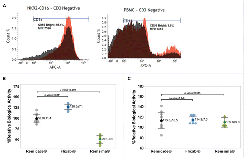
Figure 4. Correlation between percentage afucose plus percentage high mannosylated (HM) glycans and Fc functions for Remicade®, Flixabi®, and Remsima®. (A) Correlation between %afucose plus %HM glycan and %relative FcγRIIIa binding activity. (B) Correlation between %afucose plus %HM glycan and %relative ADCC activity.
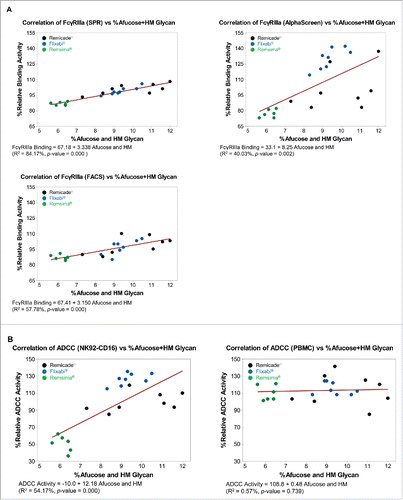
Table 1. %Afucose plus high mannosylated glycan and %relative ADCC activity of infliximab products. %RBA: %relative biological activity.
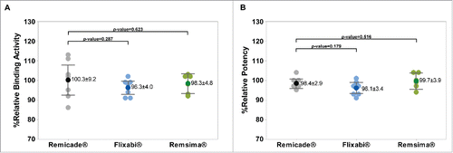
Figure 5. Fab-related TNF-α binding and TNF-α neutralization activities of Remicade®, Flixabi®, and Remsima®. (A) Percentage of relative binding activity assessed by fluorescence resonance energy transfer. (B) Percentage of relative potency measured by a luciferase reporter gene assay. Light color dots: Individual data; Dark color dots: Mean data with mean value and standard deviation; Bar: 95% confidence interval of mean.