
Figures & data
Figure 1. Composition of IgG2/4 control antibody NHDL
A, The expression of all possible VL/VH combinations for IgG2/4 control antibody generation was tested by transient transfection of HEK293E cells with different combinations of the two separate DNA plasmids encoding the light (VL+CL; pLNOk) or heavy (VH+CH; pLNOH) chains of anti-NIP (aNIP), anti-porcine CD14 (Mil2) or anti-human CD14 (18D11) specificities. Cell culture supernatant (SN) was harvested 3 days post transfection and IgG titer was estimated as µg/mL using ELISA (n = 3; mean ± S.E.M.). B, Schematic model of NHDL, a mouse/human chimeric IgG2/4 antibody. The constant regions, i.e., human kappa light chain (hCκ; blue), human IgG2 CH1/hinge (light gray) and human IgG4 CH2-3 (dark gray), are identical for the IgG2/4 CH hybrid antibodies described so far, i.e., NHDL, r18D11, rMil2, and eculizumab. The murine variable regions of NHDL are derived from two different specificities, i.e., VH of anti-NIP (yellow) and VL of anti-CD14 clone 18D11 (orange). The model was generated using Adobe Illustrator.
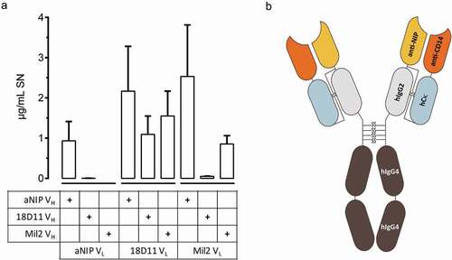
Figure 2. Structural integrity of NHDL compared to other IgG2/4 antibodies
a, The IgG2/4 antibodies NHDL, raNIP, and r18D11, all purified from the supernatant of transiently expressing HEK293E cells, as well as rMil2 and eculizumab were subjected to non-reducing SDS-PAGE (4–15%) using Precision Plus ProteinTM standard as protein ladder. b, Size exclusion chromatography profiles of the indicated IgG2/4 antibodies were normalized to a relative absorbance maximum of 1.0 and displayed as an overlay. Fractions of monomeric single IgG molecules were contained by the main peaks between 11.4 mL and 12.5 mL, while IgG oligomers or aggregates appeared earlier and as minor peaks. Note that r18D11 eluted slightly earlier than the other IgG2/4 antibodies. c, Monomeric IgG2/4 antibody fractions prepared by SEC were subjected to differential scanning fluorimetry and melting curves were generated for temperatures between 20⁰C to 80⁰C. Dye incorporation increases as proteins disassemble, reaching a maximum of around 70⁰C prior to aggregation and decrease of the signal. The derived melting temperatures were 67.7⁰C for NHDL, 64.9⁰C for r18D11, 68.5⁰C for rMil2, and 68.2⁰C for eculizumab. The melting curve of raNIP showed unusual high background fluorescence below 40⁰C, in the so-called native state.
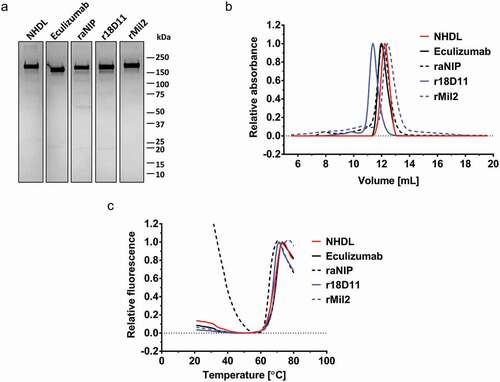
Figure 3. Test of binding and blocking activity of human CD14 by NHDL
a, Different molar ratios of purified antibody (r18D11 or NHDL) and recombinant soluble CD14 (sCD14) (given as antigen to antibody) were incubated in vitro and subsequently separated by native PAGE in absence of SDS. The antigen-antibody complex formation became apparent as a newly formed band and a reduction in or loss of the bands corresponding to free antibody or free sCD14. All three bands are indicated by arrows. b, Human whole blood was incubated with 10–10,000 ng/mL of PE-conjugated NHDL or r18D11. Binding was analyzed by flow cytometry and given as mean fluorescence intensity (MFI). For competitive binding, blood was pre-incubated with 15 µg/mL unconjugated r18D11 or NHDL prior to the addition of the PE-conjugated antibodies. Results are shown as MFI (n = 3; mean ± S.E.M.). The MFI (330 ± 9.8) of a PE-conjugated mIgG1 control antibody is indicated by the dotted line. c, Whole human blood was incubated with PBS or 100 ng/mL upLPS in absence or presence of 15 µg/mL or 30 µg/mL r18D11 or NHDL for 120 min at 37⁰C. Plasma IL-6 levels were analyzed by Bioplex technology and are given in pg/mL (n = 3; mean ± S.E.M.). d, human plasma (a pool of n = 6) was incubated with Dynabeads®-coupled NHDL, r18D11, eculizumab, or a control antibody (Ctrl IP; anti-CD3 mIgG1), and co-immunoprecipitated proteins were identified using mass spectrometry. The results are shown for non-IgG sequences with #PSM>10 and peak area values larger for NHDL than for the control antibody (Ctrl IP). The unfiltered results are displayed as a heatmap in Figure S2a.
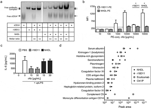
Figure 4. Test of binding and blocking of human C5 and porcine CD14 by NHDL
a, Increasing amounts (2.5–50 µg/mL) of recombinant antibody (rAb), i.e., NHDL or eculizumab, were incubated with a human serum pool that naturally contains C5, and C5-bound antibody (rAb-C5 complex) was quantified as µg/mL by ELISA according to a standard curve using eculizumab. b, Complement activation in a human serum pool in the presence or absence of 1 mg/mL heat-aggregated IgG (HAIgG) and 200 µg/mL NHDL or eculizumab (Ec.) was measured as TCC formation using ELISA. Data are given as complement arbitrary units (CAU/mL) for one representative experiment. c, Competitive binding of FITC-conjugated Mil2 to porcine whole blood cells in presence of 10, 50 or 100 µg/mL NHDL or rMil2 was measured by flow cytometry. A FITC-conjugated mIgG2b antibody served as a negative control (Ctrl). Results are shown as MFI (n = 3; mean ± S.E.M.). d, E. coli-induced (1 x 10Citation5/mL) porcine plasma IL-8 release was measured in the absence or presence of 50 or 100 µg/mL NHDL or rMil2 using Bioplex technology. The results are given as pg/mL (n = 3; mean ± S.E.M.).
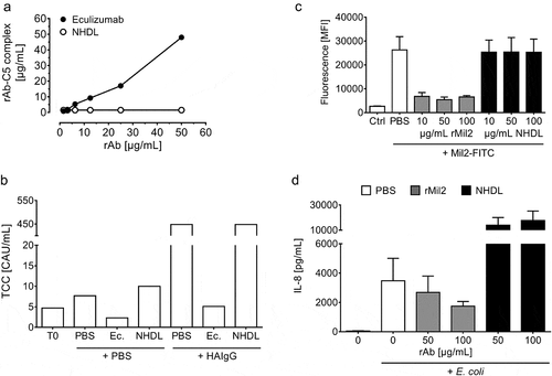
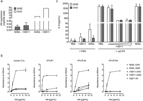
Figure 5. Characterization of CHO-produced NHDL and r18D11
A, The expression of recombinant NHDL and r18D11 by stably transfected CHO-K1SP cell pools (fed-batch) was compared to transient expression by HEK293E cells (batch-batch). Cell culture supernatants were analyzed for IgG levels using ELISA. Results are given as single value mg IgG per E06 CHO cells (n = 1), and mean mg IgG ± S.E.M. per E06 HEK293E cells for NHDL (n = 3) and r18D11 (n = 6). B, Binding of increasing concentrations of CHO- and HEK293E-produced NHDL or r18D11 to recombinant human C1q or recombinant human Fcγ receptors hFcγRI, hFcγRIIa, and hFcγRIIIa at pH 7.4 was measured by ELISA. As a positive control, a recombinant human IgG1 antibody (bevacizumab (Avastin®), Roche) was included in the analysis. The results are shown as relative units for the absorbance at 450 nm (n = 2; mean ± S.D.). C, Human whole blood was incubated with CHO- or HEK293E-produced r18D11 (1.5, 15 and 30 µg/mL) or NHDL (15 µg/mL) in the presence of 100 ng/mL upLPS. As controls, 15 µg/mL r18D11 or NHDL were incubated in the presence of PBS, only. Plasma IL-6 was measured using Bioplex technology. Data are given as pg/mL (n = 2; mean ± S.D.).
Supplemental material