Figures & data
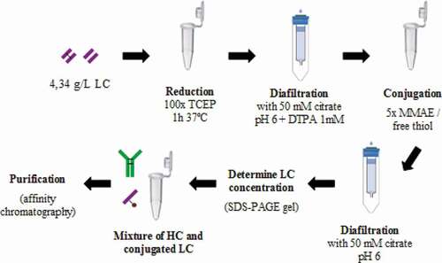
Figure 1. Strategy to obtain DAR 2 homogeneous ADCs. anti-HER2 chains were independently produced in recombinant HEK293 cultures. Then, each chain was purified by affinity chromatography and light chain (LC) was conjugated to vcMMAE. Finally, the complete mAb was assembled
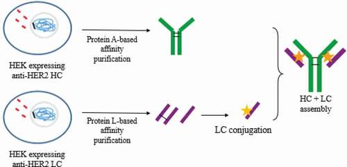
Figure 2. HC and LC conformation of proteins expressed by recombinant HEK293. (a) SDS-PAGE gel (denaturing conditions). (b) SEC-HPLC (native conditions) for HC. (c) SEC-HPLC for LC
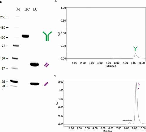
Table 1. Molecules detected under native conditions (SEC-HPLC)
Table 2. Isolated antigen HER2 recognition in the ELISA test
Figure 3. HIC-HPLC chromatogram. LC (dotted line) appears in two peaks, corresponding to the (un)covalently bonded LC dimers. LC-MMAE (continuous line) appears as a single peak at 8.1 minutes
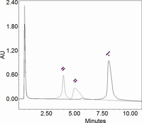
Figure 4. Comparison of HICxSEC-native MS chromatograms of ADC and naked mAb. a) DAR 2 HIC profile. b) DAR 2 deconvoluted spectra. c) Naked mAb HIC profile. d) Naked mAb deconvoluted spectra
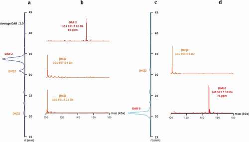
Figure 5. MS and MS/MS spectra of the two peptides containing the MMAE payload linked to the Cys214 localized on the LC part of the ADC. (a) [SFNRGEC] + 1 payload, (b) [GEC] + 1 payload
![Figure 5. MS and MS/MS spectra of the two peptides containing the MMAE payload linked to the Cys214 localized on the LC part of the ADC. (a) [SFNRGEC] + 1 payload, (b) [GEC] + 1 payload](/cms/asset/971db405-6bf8-49f6-a63b-7e4e8a5360b4/kmab_a_1702262_f0005_oc.jpg)
Figure 6. Biological activity of the in vitro assembled mAb and homogeneous DAR 2 ADC compared to in vivo folded mAb and heterogeneous DAR 4 ADC, respectively. Cell inhibition assay. Orange: DAR 2 homogeneous T-MMAE assembled by the strategy described. Black: assembled non-conjugated mAb by the strategy described. Green: DAR 4 heterogeneous T-MMAE (reference). Blue: in vivo folded trastuzumab. The concentration indicated is referred to the complete ADC (mainly mAb) and not to toxin:payload
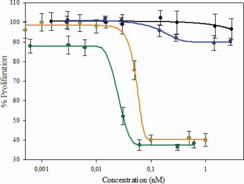
Figure 7. Future proposed strategies based on the described method to obtain homogeneous ADCs. The same conjugation could occur in either LC or HC. After assembly, the ADC could be loaded with 2 different site-directed payloads
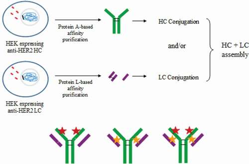
Figure 8. Reduction-oxidation assembly approach. In vivo produced and purified LC and HC are reduced to free disulfide bonds and deoxidized to obtain the assembled mAb
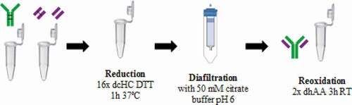
Figure 9. Spontaneous assembly approach to generate homogeneous ADC. LC are reduced to free interchain cysteines and allow the site-directed payload:linker conjugation. Then, dcHC is mixed to obtain the assembled ADC