
Figures & data
Table 1. Serum titer analysis for immunized animals by FACS
Table 2. VHH competition with fractalkine
Table 3. Percent block of fractalkine-induced chemotaxis by VHHs
Table 4. Functional profiling of Bi-valent VHHs
Figure 1. Binding of bivalent family101 VHHs to Ba/F3/hCX3CR1 cells. Mean fluorescence per cell was measured by flow cytometry of various concentrations of Alexa Fluor labeled VHHs. IC50s were calculated to be <1 nM for all four constructs tested
Table 5. Comparison between wildtype and sequence optimized VHH
Table 6. Functional profile of BI 655088 and BI 655089
Figure 2. Exemplar Chemotaxis Assay data showing the functional activity of BI 655088
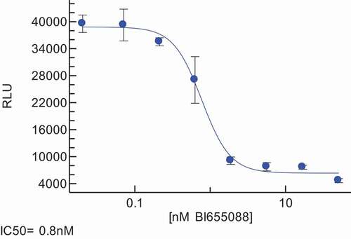
Table 7. Pharmacokinetic profile of BI 655088 and BI 655089
Figure 3. Characterization of Target Engagement Biomarkers in hCX3CR1 KI Mouse Model. Free receptor levels, serum concentrations of BI 655088 and serum fractalkine levels in hCX3CR1 KI mice after receiving two doses at 30 mg/kg of BI 655088 or vehicle. Day 0 represents baseline concentrations for free receptor and serum fractalkine of hCX3CR1 KI mice dosed with vehicle control (Mean ± SD, *p < .0001 vs. Day 0 and Day 13). BI 655088 and Fractalkine levels were determined by immunoassay and free receptor levels assessed by FACS
Figure 4. Human CX3CR1 is expressed in the atherosclerotic plaques of hCX3CR1/ApoE KO mice (brachiocephalic artery) after feeding with an HF/HC diet (a). IHC was done using a human anti-CX3CR1 antibody and compared to a no-antibody negative control. Similar expression of CX3CR1 was also seen in the plaque from a cross-section of human coronary artery from an atherosclerotic patient (b)
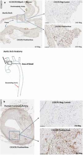
Figure 5. BI 665088 treatment significantly reduced the descending aorta plaque area (a) with a concomitant increase in serum fractalkine (b). Representative images of descending aortas after Oil Red O staining for plaque area (c)
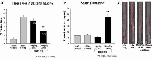
Table 8. Developability of BI 655088