
Figures & data
Figure 1. Workflow for anti-BDNF antibody affinity maturation
A: The CDR-grafted variant of chicken R3bH01, H01, was chosen as a template for affinity optimization using a soft mutagenesis library-based approach. Mutagenic libraries of H01 were designed as single-chain variable fragments (scFvs) were subjected to soft-randomization across VH CDR1, VH CDR2, VH CDR3, VLCDR1 and VL CDR3 using a series of mutagenic primers as shown. A stepwise library selection approach was taken incorporating off-rate challenge with 100-fold competing BDNF for increasing times over each round and outputs were interrogated by high throughput screening by competition ELISA. Clones demonstrating beneficial CDR-based mutations compared to the parental were combined at the IgG1 reformatting stage. The clone with the best characteristics, B30, was chosen as the lead with a KinExa-measured affinity of approximately 1 pM.
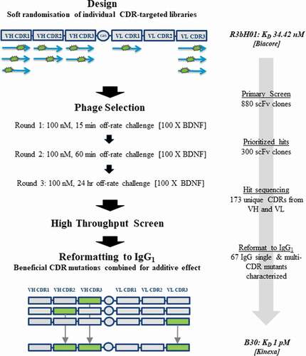
Figure 2. High-throughput screening of soft mutagenesis phage libraries for affinity-improved anti-BDNF antibodies
A: A high-throughput single-point periplasmic competition HTRF assay was used to identify clones with potential improvements in binding over the parental chimeric R3bH01 and humanized H01 in an scFv format. Each point (Red: Round 1 clones, Blue: Round 2 clones, Yellow: Round 3 clones) represents a selection output clone. The data is represented as % inhibition of R3bH01-BDNF interaction compared to the negative control (turquoise, unrelated scFv). Humanized H01 (green) and chimeric R3bH01 (black) as scFv are shown for comparison. B: Prioritized mutants were reformatted to IgG1 and ranked for potency using a functional pERK signaling assay. R3bH01 (green) is highlighted and compared to B30 (blue). Data was plotted as relative response units versus log molar concentration of purified IgG1.
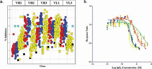
Table 1. Comparison of parental chicken R3bH01 to humanized & affinity optimized B30
Figure 3. Demonstration of affinity improvement of B30 over parental R3bH01
Human BDNF was directly immobilized on a CM5 Biacore sensor chip surface. A dilution series of parental antibody R3bH01 (A) and the affinity optimized antibody B30 (B) were injected, allowed to associate, followed by dissociation in HBS-EP+ running buffer. All data were fit to a 1:1 Langmuir binding model using T200 evaluation software v1.0. Data shown are representative curve fits for at least 2 independent dilution series experiments. Biacore measurements shown were performed at 37ºC. C: Binding of Fab B30 to Human BDNF by Kinetic Exclusion Assay. The B30 Fab, ranging in concentration from 2 nM to 343 fM was mixed with a fixed concentration of human BDNF, either at 60 pM for a KD controlled curve or 400 pM for a receptor-controlled curve. All solutions of Fab and antigen were incubated overnight at room temperature to attain equilibrium. Data points are a measurement of percentage free human BDNF at each concentration of B30 Fab. D: N-curve analysis was used to globally fit the curve data to obtain an equilibrium dissociation constant, or KD value.
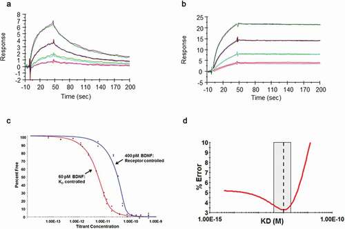
Figure 4. Affinity of anti-BDNF antibodies correlates to potency of neuropathy-induced Kv current suppression
A: Representative voltage-clamp traces from acutely isolated DRG neuron from rats dosed with an isotype control antibody (0.1 mg/kg). Conductances from native Kv ion channels in an uninjured DRG neuron (black traces) are much larger than those from an injured DRG neuron (red traces). Quantification of native potassium conductance at the end of the voltage step (often termed Ik) is plotted as an I/V curve (inset shows area under the curve analysis (pA.mV.pF−Citation1). All subsequent data is analyzed in this way with statistics being analyzed from the AUC analysis. B: Representative voltage-clamp traces from acutely isolated DRG neuron from rats dosed with a neutralizing anti-BDNF antibody (B30 0.1 mg/kg). The suppression of Kv ion channels is completely reversed by neutralization of BDNF so that the injured and uninjured conductances (and AUC values) are indistinguishable. C: R3bH01 reverses injury-induced Kv suppression in a dose-dependent manner. Voltage-activated potassium currents (as shown in A and B) are represented as a function of voltage. DRG neurons from isotype control-treated animals exhibited significantly different Kv currents as did those from animals treated with 0.1 mg/kg R3bH01. Injured DRG neurons from animals dosed with 1 or 10 mg/kg R3bH01 exhibited no significant difference in Kv currents when compared to uninjured neurons. Data are represented as mean values ± SEMs, data are analyzed using area under the curve analysis (as in A and B), unpaired t-tests were utilized to assess significance (ns = not significant, *** = p < .001). D: B30 reverses injury-induced Kv suppression in a highly potent dose-dependent manner. DRG neurons from isotype control-treated animals exhibited significantly different Kv currents as did those from animals treated with 0.01 mg/kg. Injured DRG neurons from animals dosed with 0.03 or 0.1 mg/kg B30 exhibited no significant difference in Kv currents when compared to uninjured neurons. Data are represented as mean values ± SEMs, data are analyzed using area under the curve analysis (as in A and B), unpaired t-tests were utilized to assess significance (ns = not significant, *** = p < .001).
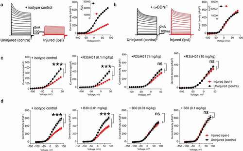
Figure 5. B30 reduces thermal hyperalgesia in the tibial nerve of SNL injured rats
Multi-unit extracellular recordings were made of basal nerve activity as well as thermally evoked afferent nerve firing from the tibial nerve of neuropathic rats 2–3 weeks post SNL injury and dosed for 4–7 days with hIgG1 or the anti BDNF antibody (B30, 0.03 or 0.1mpk). Representative trace demonstrating robust afferent nerve firing in response to a non-noxious heat ramp (36-48°C, at 0.2°C/s; A), as well as high levels of basal nerve activity (highlighted by oval in each trace) in SNL injured animals dosed with hIgG1 (B). Animals dosed with B30 at 0.03 mpk (C) exhibited similar levels of basal nerve activity but reduced thermally evoked afferent firing. Animals dosed with B30 at 0.1mpk (D) exhibited reduced basal nerve firing and minimal thermally evoked afferent firing. In all cases nerve activity was fully inhibited by 1 mM lidocaine. B30 at 0.03 and 0.1 mpk significantly inhibits injury induced firing to a non-noxious heat ramp in relation to hIgG1 dosed animals (E). B30 at 0.1mpk also significantly attenuates afferent firing elicited by a noxious heat ramp (36-52°C, at 0.4°C/s; E) as well as basal nerve firing (F). Data is represented as mean values ± SEMs, data were analyzed with single parametric analysis and compared to isotype control (* = p < .05, ** = p < .01, *** = p < .001).
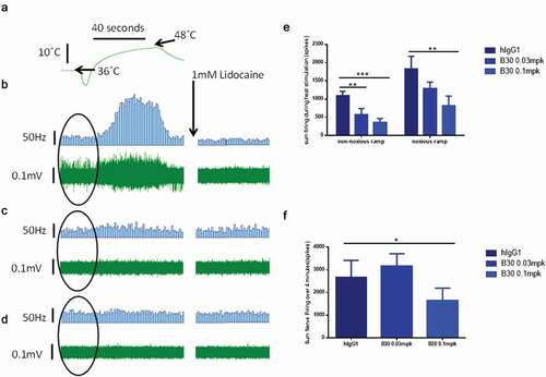
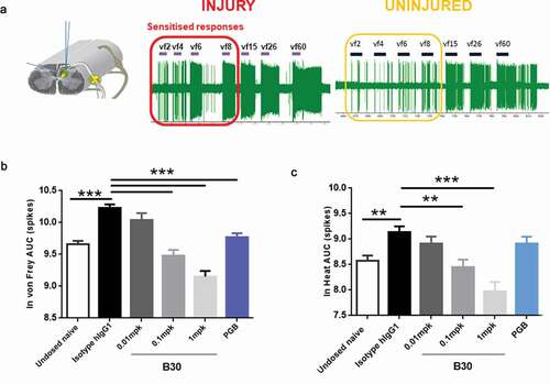
Figure 6. B30 reduces spinal cord hyperexcitability in spinal dorsal horn (DH) neurones of nerve injured animals
A: Recordings made from ipsilateral DH neurones 3 weeks after nerve injury show exaggerated firing response to mechanical punctate stimuli. This is particularly prominent with von frey stimuli in the low intensity range (2 to 8 grams). B and C. Spinal cord recordings were made in neuropathic rats, 4–7 days after being dosed with hIgG1 control, B30 (0.01, 0.1 and 1 mg/kg) or pregabalin. In addition, a separate group of undosed naïve animals were included in the study. Graded mechanical and heat stimuli were applied to the central receptive field and the AUC of the stimulus response curve was quantified. Mechanical (B) and heat hyperexcitability (C) induced by peripheral nerve injury was dose dependently reduced by the anti-BDNF antibody, B30. Pregabalin treatment over 5 days (15 mg/kg, po) produced attenuation of the mechanical evoked responses, but not responses to heat. **p < .01; p < .001 compared to hIgG1 negative control.
Supplemental material