Figures & data
Figure 1. Characterization of CLEC5A expression on primary myeloid cells (a) FACS plots showing expression of CLEC5A on human primary monocyte-derived macrophages, monocytes, and neutrophils. (b) FACS plots showing expression of CLEC5A on mouse BMDMs and peritoneal macrophages. (c and d) Effect of human and mouse macrophage polarization states on CLEC5A expression. (A) Three panels of FACS histogram of CLEC5A expression on human macrophages (left panel, high signal), monocytes (central panel, moderate signal), and neutrophils (right panel, minimal signal). (B) Two panels of FACS histogram of CLEC5A expression on murine BMDMs (left panel), and murine peritoneal macrophages (right panel). (c). Bar graph with individual bars representing mean fluorescence intensity (MFI) of anti-CLEC5A staining on human macrophages with various treatments. (d). Bar graph with individual bars representing mean fluorescence intensity (MFI) of anti-CLEC5A staining on murine BMDMs with various treatments.
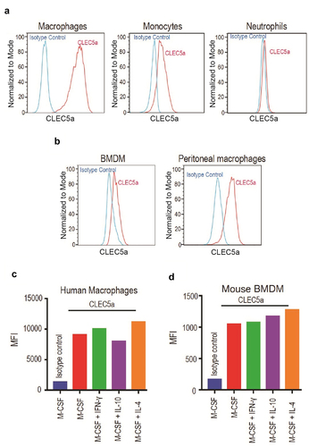
Figure 2. Characterization of CLEC5A expression on mouse tumor infiltrating cells CLEC5A expression on different cell types in MC38 and CT26 tumors (Mean ± S.D of MFI from 3 independent tumors).
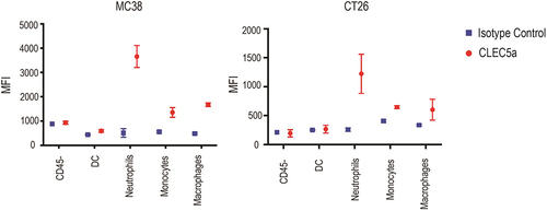
Figure 3. Identification and characterization of CLEC5A agonist antibodies CLEC5A agonist antibody induced TNF production from CLEC5A + human primary macrophages (Mean ± S.D of triplicates from one donor). (b) Schematic depiction of B cell-dependent engagement of CLEC5A via CD20/CLEC5A bispecific antibody. (c) Schematic depiction of different CLEC5A bispecific antibodies. Bispecific antibodies carry LALAPG mutation in their Fc while CD20 hIgG1 has a wild-type Fc. (d) Binding Kd values of CLEC5A bispecific antibodies for human and mouse CLEC5A were determined by Biacore. (e) Activation of CLEC5A signaling (TNF production) in human macrophages by the bispecific antibody in the presence of target Raji cells (Mean ± S.D of triplicates from one donor).
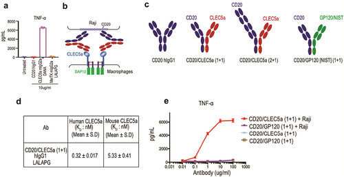
Figure 4. Characterizing activity and potency of CLEC5A-dependent bispecific antibodies (a and b) Raji cell killing by human macrophages in the presence of indicated bispecific antibody and blocking agents (Mean ± S.D of triplicates from one donor, Effector: Target ratio is 5:1). (c) CD20/CLEC5A-dependent human macrophage-mediated cell-killing is specific to Raji (CD20+) but not to Jurkat (CD20−) (Mean ± S.D of triplicates from one donor, Effector: Target ratio is 5:1). (d) EC50 of macrophage-mediated tumor (Raji) cell-killing in the presence of indicated antibodies. Data were derived from 4 independent human donors. Effector: Target ratio is 5:1. (e) IncuCyte phagocytosis assay demonstrates the uptake of pHrodo-labeled Raji cells by the human macrophages in the presence of indicated antibodies. Antibody concentration, 2ug/ml. Effector: Target ratio is 1:5. (f) Testing the dual action of anti-CD20 and CLEC5A bispecific antibody in human macrophage-mediated Raji cell killing (Mean ± S.D of triplicates from one donor, Effector: Target ratio is 5:1).
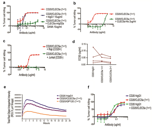
Figure 5. Demonstration of CLEC5A-dependent phagocytosis of solid tumor cells Activation of CLEC5A (TNF production) by the bispecific antibody in the presence of target SK-BR-3 cells (Mean ± S.D of triplicates from one donor). (b) Macrophage-mediated tumor (SK-BR-3) cell killing in the presence of indicated bispecific antibody (Mean ± S.D of triplicates from one donor). Effector: Target ratio for phagocytosis and cytokine assay is 5:1.
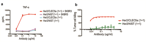
Figure 6. Characterization of cross-species agonistic activity of CLEC5A bispecific antibody Activation of CLEC5A (TNF production) in mouse BMDMs by the bispecific antibody in the presence of target Raji cells (Mean ± S.D of triplicates). (b) Mouse BMDM-mediated tumor (Raji) cell-killing in the presence of indicated bispecific antibodies (Mean ± S.D of triplicates). Effector: Target ratio for phagocytosis and cytokine assays is 5:1.
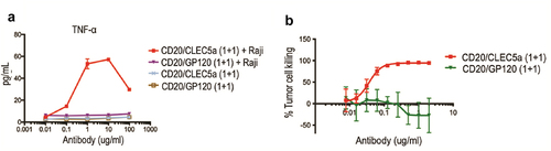
Figure 7. Characterization of the OX40/CLEC5A bispecific antibodies FACS plots depicting stable expression of mOX40 on EL4 cells (left panel). (b) Human macrophage-mediated EL4-mOX40 cell-killing in the presence of indicated bispecific antibody (Mean ± S.D of triplicates from one donor, Effector: Target ratio is 5:1).
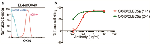
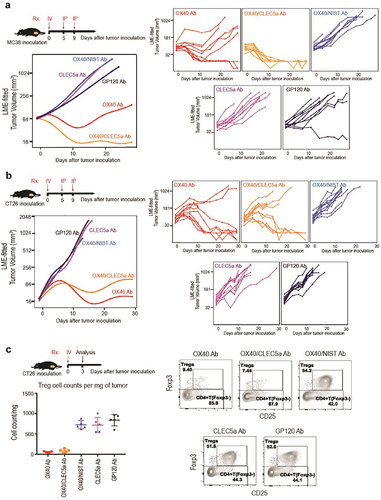
Figure 8. Depletion of Tumor-infiltrating regulatory T cells by the OX40/CLEC5A bispecific antibody results in antitumor efficacy (a and b) Growth of MC38 and CT26 tumors in mice treated with the indicated antibodies (n = 10 mice/group). Treatment started 9 days after tumor cell inoculation. (c) Quantification of CD4+ FoXP3+ CD25+ tumor Treg cells after treatment with indicated antibodies (Left panel). Representative FACS plots showing the percentage of Treg cells within total CD4+ T cells in tumors with different treatments (Right panel). Mice were treated with indicated antibodies (n = 5 mice/group) on day 9 after CT26 tumor inoculation, and tumors were harvested on day 3 after antibody treatment. Antibodies used: OX40 mIgG2a (OX40 Ab), OX40/CLEC5A mIgG2a LALAPG (OX40/CLEC5A Ab), OX40/NIST mIgG2a LALAPG (OX40/NIST Ab), CLEC5A mIgG2a DANA (CLEC5A Ab), and GP120 mIgG2a LALAPG (GP120 Ab).