Figures & data
Figure 1. Engineering novel N-glycan sites into the F5111 antibody. (a) Crystallographic structure of the heavy chain (HC) and light chain (LC) variable domains of the F5111 antibody in complex with the IL-2 cytokine (PDB ID: 5UTZ), overlaid with the heterotrimeric IL-2 receptor (PDB ID: 2B5I). Complementarity-determining loops of the F5111 HC (H1, H2, and H3) and LC (L1, L2, and L3) are indicated (inset). Green spheres indicate the location of the asparagine residue in the engineered N-glycan site. (b) Reducing SDS-PAGE analysis of six glycovariants of the F5111 antibody with inserted N-linked glycosylation sites in the HC (M1, M2, and M3) or LC (M4, M5, and M6). (c) Con-A lectin blot demonstrating glycosylation of F5111 variants. WT, wild type.
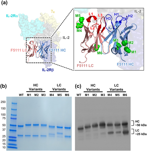
Figure 2. F5111 glycovariants show similar binding and functional properties to parent antibody. (a) BLI studies depicting the interaction between immobilized human IL-2 and soluble antibody (either F5111 or glycovariants thereof). (b, c) Competitive BLI-based IL-2 binding studies between F5111 or glycovariants thereof and IL-2 receptor subunits. Equilibrium binding of a saturating concentration of IL-2 (600 nM) to immobilized IL-2Rα (b) or IL-2Rβ (c) in the presence of titrated amounts of F5111 antibody. (d, e) IL-2 signaling pathway activation induced by a 1:1 molar ratio of F5111:IL-2 complexes on human YT-1 human NK cells with (d) and without (e) IL-2Rα expression. STAT5 phosphorylation was detected via flow cytometry. Error bars represent standard deviation. (f) Comparison of YT-:YT+ EC50 values for signaling activation by IL-2 or various IL-2/antibody complexes. WT, wild type.
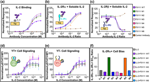
Figure 3. Engineering novel N-linked glycosylation sites into the 602 antibody. (a) Threaded computational model of the heavy chain (HC) and light chain (LC) variable domains of the 602 antibody in complex with the IL-2 cytokine (generated using the iTASSER server), overlaid with the heterotrimeric IL-2 receptor (PDB ID: 2B5I). Complementarity-determining loops of the HC (H1, H2, and H3) and LC (L1, L2, and L3) are indicated (inset). Green spheres indicate the location of the asparagine residue in the engineered N-glycan site. (b) Reducing SDS-PAGE analysis of 6 glycovariants of the 602 antibody with inserted N-linked glycosylation sites in the HC (M1 and M9) or LC (M2, M4, M8). (c) Con-A lectin blot demonstrating glycosylation of 602 variants. Glycophorin A (GlyA) and ovalbumin (OVA) were used as controls for O-linked and N-linked glycosylation, respectively. WT, wild type.
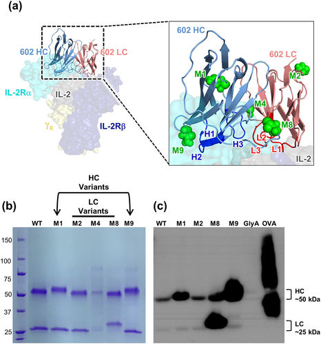
Figure 4. 602 glycovariants recapitulate binding and functional properties of parent antibody. (a) BLI studies depicting the interaction between immobilized human IL-2 and soluble antibody (either 602 or glycovariants thereof). (b, c) BLI-based competitive IL-2 binding studies between 602 or glycovariants thereof and IL-2 receptor subunits. Equilibrium binding of a saturating concentration of IL-2 (600 nM) to immobilized IL-2Rα (b) or IL-2Rβ (c) in the presence of titrated amounts of 602 antibody is shown. (d, e) IL-2 signaling pathway activation induced by a 1:1 molar ratio of 602:IL-2 complexes on human YT-1 human NK cells with (d) and without (e) IL-2Rα expression. STAT5 phosphorylation was detected via flow cytometry. Error bars represent standard deviation. (f) Ratio of YT-:YT+ EC50 values for signaling activation by IL-2 or various IL-2/antibody complexes. WT, wild type.

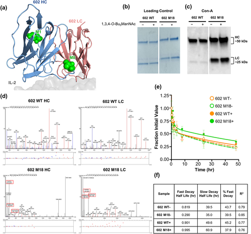
Figure 5. Glycosylation and pharmacokinetic properties of the M18 double gain-of-glycan 602 variant. (a) Threaded computational model of the heavy chain (HC) and light chain (LC) variable domains of the 602 antibody in complex with IL-2 (generated using the iTASSER server). The two installed glycosylation sites for the M18 variant are indicated as green spheres. (b,c) Reducing SDS-PAGE analysis (b) and ConA lectin immunoblot (c) showing glycosylation of the wild-type 602 antibody compared to the M18 variant. Proteins were expressed in HEK293 F (HEK) or CHO-S (CHO) cells, as indicated, in the absence (-) or presence (+) of 1,3,4-O-Bu3ManNAc. (d) Fragmentation spectra of wild-type and corresponding engineered N-linked glycovariant peptides from the heavy and light chains of both 602 wild-type and 602 M18, respectively. Oxonium ions from 602 M18 glycopeptides are boxed in red. (e) In vivo pharmacokinetic study of the parent 602 antibody versus the M18 variant, prepared with or without 1,3,4-O-Bu3ManNAc. Serum concentration is plotted at various time points following r.o. injection (n = 3). WT, wild type.