Figures & data
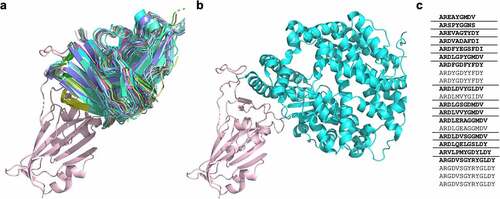
Figure 1. a) A cartoon representation of 22 antibodies binding to the “neck” of SARS-CoV-2 RBD (salmon), with b) showing ACE-2 (turquoise) binding at the same site. c) The CDRH3 sequences represented across the 22 RBD ‘neck’-binding antibodies. Lenient VH-clonotypes are separated with solid lines, with the cluster representative highlighted in bold font (derived from Robinson et al.Citation39).