
Figures & data
Figure 1. Binding epitopes of antigen-antibody pairs as determined by different technologies.
The amino acids forming the binding epitopes of the antigen–antibody pairs, as identified by multiple epitope mapping technologies are highlighted on model structures of the antigens and their amino acid sequences. a) PD-1+pembrolizumab. b) PD-1+nivolumab. c) CTLA-4+ipilimumab. d) CTLA-4+tremelimumab. e) CD27+MK-5890. X-ray: X-ray crystallography. PepArr: Peptide array. ALN: Alanine scanning; red residues resulted in loss of binding upon mutation while gray residues were mutated and showed negative result in ALN. DomX: Domain exchange. HDX: Hydrogen-deuterium exchange. XL: Chemical cross-linking. HRF: Hydroxyl Radical Footprinting.
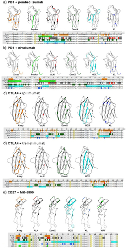
Figure 2. Antibody binding to mouse/human domain exchange mutants.
Mouse/human exchange mutants were transiently transfected into CHO-K1 cells, after which antibody binding was determined. a) Binding of pembrolizumab and nivolumab to mouse/human PD-1 domain exchange mutants was determined by cell ELISA (left). The designs of the mutants are shown on the right. Purple represents human domains and green represents mouse domains. b) Binding of ipilimumab and tremelimumab to human CTLA-4, mouse CTLA-4 and CTLA-4 mouse/human exchange mutants was determined by cell ELISA. In ribbon drawings, green represents human domain and brown represents mouse domain. c) Binding of MK-5890 to CD27 mouse/human domain exchange mutants, as established by FACS. Human CRDs are indicated in blue, mouse CRDs in red.
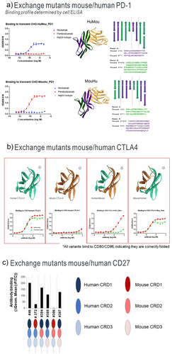
Figure 3. Binding of anti-CTLA-4 antibodies to alanine mutants.
Single amino acids of CTLA-4 were mutated into alanine and mutant proteins were expressed on CHO-K1 cells. Binding of ipilimumab, tremelimumab and the CTLA-4 ligands CD80 and CD86 is expressed relative to antibody binding to wild type hCTLA-4 (hCTLA-4WT), which was set at 100%. Amino acid positions of the alanine substitutions are indicated.
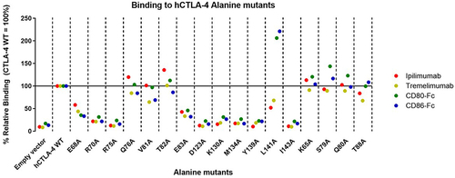
Table 1. Pro and cons of each technique.
Supplemental material