Figures & data
Figure 1. Venn diagram of Plasmodium falciparum 3D7 proteins reported in extracellular vesicles during parasite infection according to several authors
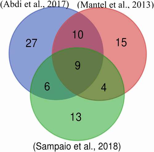
Table 1. Dali best results for P. falciparum molecular mimicry candidates vs human proteins
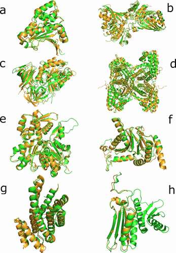
Figure 2. Superimposition of candidate proteins. (a) Heat shock protein 90, 6CEO (Homo sapiens) with 3K60 (P. falciparum). (b) Glyceraldehyde-3-phosphate dehydrogenase, 3GPD (Homo sapiens) with 1YWG (P. falciparum). (c) Elongation factor 1-alpha, 3C5J (Homo sapiens) with the homology predicted structure for (P. falciparum). (d) Fructose-aldose bisphosphate, 5LN3 (Homo sapiens) with 6MUX (P. falciparum). (e) Actin-1, 5 JLH (Homo sapiens) with 6I4K (P. falciparum). (f) Proteasome subunit alpha, 6REY (Homo sapiens) with 6MUW (P. falciparum). (g) 14-3-3 proteins, 3UVW (Homo sapiens) with the homology predicted structure for P. falciparum. (h) 40S ribosomal protein S3, 6YBS chain K (Homo sapiens) with 6OKK chain D (P. falciparum). The human proteins are represented in green and the P. falciparum proteins in light brown