Figures & data
Figure 1. Simplified representation of the PBPK model simulating the absorption/metabolism/elimination (solid grey), blood distribution (solid black), direct uptake from liver through lipid deposition (dotted black), and passive diffusion in tissues (dotted grey) of α-HBCDD in growing broilers.
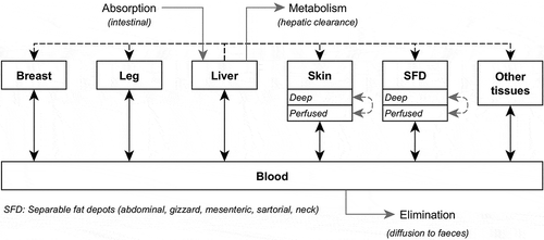
Table 1. Parameters of the model for fast-(FG) and slow-(SG) growing broilers. In brackets, parameter values after optimisation.
Table 2. Fast- (FG) and slow-growing (SG) broilers exposed to α-HBCDD through a contaminated feed (38 µg α-HBCDD kg−1) from hatching to slaughter: main characteristics of broilers, assimilation efficiency (AE) and accumulation ratio (AR) of α-HBCDD at slaughter.
Figure 2. Observed and simulated α-HBCDD concentration (ng g−1 lipid weight)1 and enantiomeric fraction (EF)2 in tissues of broilers exposed to a contaminated feed (38 µg α-HBCDD kg−1). (a): slow-growing broilers exposed during 42 days and then decontaminated during 42 days (used for optimisation); (b): slow-growing broilers exposed during 84 days; (c): fast-growing broilers exposed during 42 days.
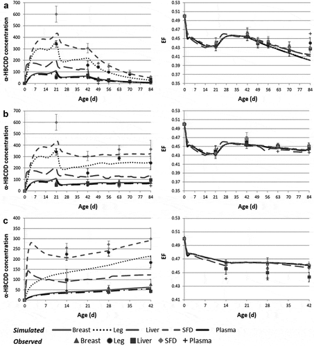
Figure 3. Simulated kinetics of α-HBCDD accumulation ratio (ARlip)1 in tissues of fast- (FG) and slow-growing (SG) broilers2 exposed through feed (38 µg α-HBCDD kg−1) from hatching to slaughter.
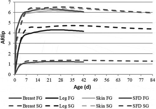
Figure 4. Assimilation efficiency (AE)1 and accumulation ratio (AR)2 of α-HBCDD in tissues of fast- (FG) and slow-growing (SG) broilers3 exposed through feed (38 µg α-HBCDD kg−1) from hatching to slaughter according to their age (Age, d) and body weight (BW, kg) at slaughter.
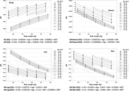
Table 3. Final concentrations, mean decontamination and elimination rates of α-HBCDD in tissues of fast- (FG) and slow-growing (SG) broilersa contaminated either through feedb or extruded polystyrenec (XPS) consumption.
Figure 5. Simulated kinetics of α-HBCDD burden (µg) (a) and accumulation ratio (ARlip)1 (b) in tissues of fast- (FG) and slow-growing (SG) broilers2 exposed through feed (38 µg α-HBCDD kg−1) from hatching to 21 and 42 days, respectively. Mean decontamination (kc) and elimination (kw) rates are presented in .
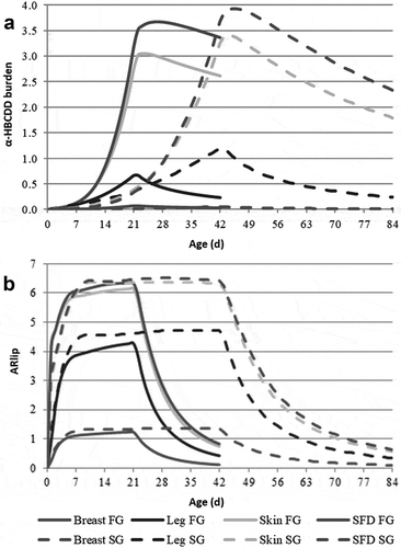
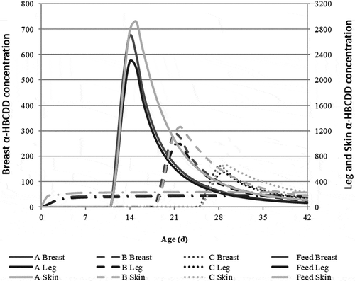
Figure 6. Simulated kinetics of α-HBCDD concentration (µg kg−1 lw) in breast, leg and skin of fast-growing broilers1 ingesting 172 µg α-HBCDD according to different modalities of exposure: 42-day exposure through feed containing 38 α-HBCDD µg kg−1 (Feed) or exposure through the daily ingestion of 57.3 µg α-HBCDD (extruded polystyrene) during 3 days from 12 to 14 (a), 19 to 21 (b) or 26 to 28 (c) days of age2,3.