
Figures & data
Table 1. Animal grouping.
Table 2. Conversion dose in mice based on body surface area (The dose used in mice was calculated by multiplying human dose by 12.3 (Food and Drug Administration, Citation2005)).
Figure 1. Immune organ index. Body weight, spleen weight, and thymus weight of mice were weighed. The spleen index (a) and thymus index (b) were calculated using the following formula: spleen index = spleen weight (milligrams)/body weight (kilograms); thymus index = thymus weight (milligrams)/body weight (kilograms).
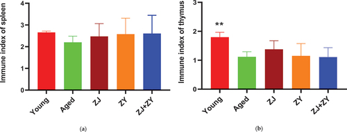
Figure 2. Lymphocyte proliferation in the spleen. Lymphocyte proliferation was detected following the preparation of splenic lymphocytes using the MTT assay. The proliferation ability was expressed by using the stimulus index (Si=absorbance of sample well/Absorbance of control well).
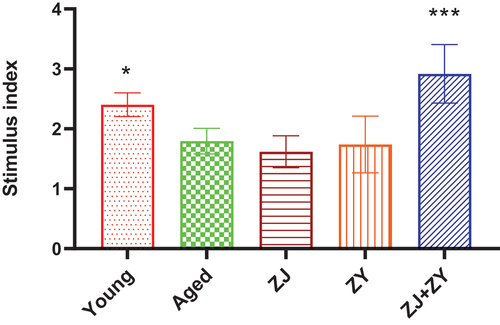
Figure 3. Lytic activity of splenic NK cells. NK cells in the spleen (effector cells) were co-cultured with YAC-1 (target cells) for 4 h. Cytotoxicity of NK cells was measured and calculated as follows: Cytotoxicity (%) = (Absorbance of sample well − Absorbance of natural release well/(Absorbance of maximum release well − Absorbance of natural release well). Note: compared with the aged group. *: P < .05, ****: P < .0001.
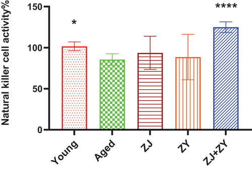
Figure 4. Cytokine secretion in serum of mice. Serum was collected following 5-week intragastric administration. Pro-inflammatory cytokines, including IL-1α, IL-1β, IL-2, IL-6, TNF-α, INF-γ, and GM-CSF (a–g), and anti-inflammatory cytokines, including IL-4, IL-5, and IL-10 (h–j), related to aging were detected.
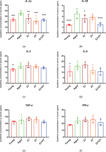
Figure 4. (Continued).
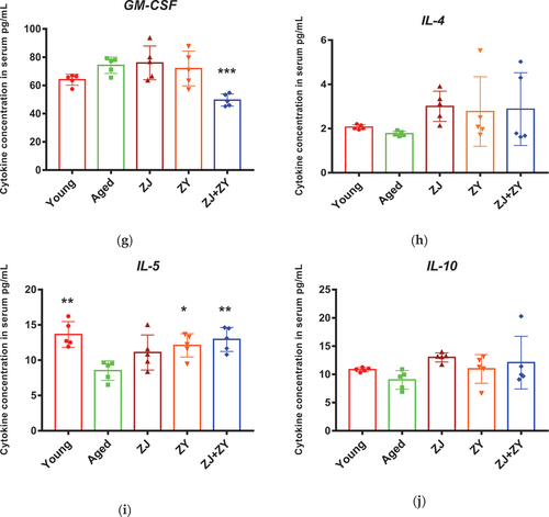
Table 3. Summary of cytokine secretion in food supplement product – treated aged mice.
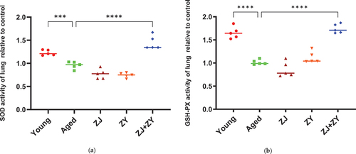
Figure 5. Antioxidant enzyme activity in lung tissue of mice. The lungs of mice were collected after 5-week intragastric administration. SOD (a) and GSH-Px (b) enzyme activities were detected using the superoxide dismutase (SOD) and glutathione peroxidase (GSH-Px) kit, respectively. Note: compared with the aged group.***: 0.0001 < P < .001, ****: P < .0001.
Data availability statement
The data in this study are available on request from the author.