Figures & data
Figure 1. Effects of xylitol on food intake (A) and body weight gain (B) in different animal groups during the whole feeding period. Fasting blood glucose concentrations were measured during the 5-week experimental period (C). Oral glucose tolerance test (OGTT) on the last day of the experimental period (D). Results are given as means ± SEM. Differences between groups were determined by ANOVA followed by Duncan’s test. * p < .05 vs control group; # p < .05 vs diabetic control group. C: control group; CD: diabetic control group; X1: 1.25 g/kg·bw xylitol; X2: 2.5 g/kg·bw xylitol; X3: 5 g/kg·bw xylitol.
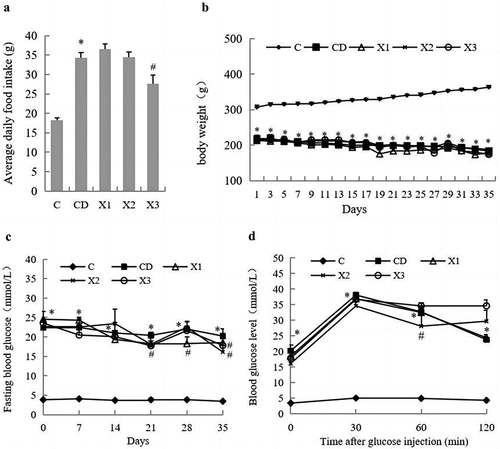
Figure 2. Xylitol had no significant effect on insulin concentration in serum but ameliorated lipid disorders in STZ-induced diabetic rats. (A) serum insulin level, (B) TG: triglyceride, (C) TC: total cholesterol, (D) HDL-C: high density lipoprotein cholesterol. Results are given as means ± SEM. Differences between groups were determined by ANOVA followed by Duncan’s test. * p < .05 vs control group; # p < .05 vs diabetic control group. C: control group; CD: diabetic control group; X1: 1.25 g/kg·bw xylitol; X2: 2.5 g/kg·bw xylitol; X3: 5 g/kg·bw xylitol.
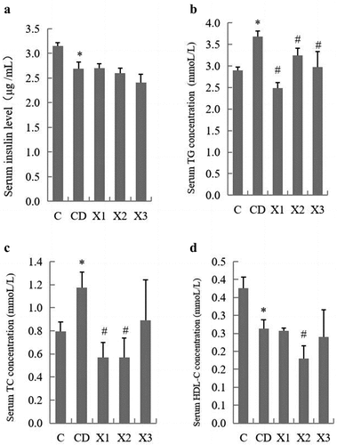
Figure 3. Xylitol ameliorated clinical symptoms in the diabetic rat in small intestine. (A) and (B) are representative H&E stained histological section from the jejunum and ileum (original magnifications, ×100). (C) Xylitol reduced DAO (diamine oxidase) concentration in serum of the diabetic rat. (D) Xylitol increased concentrations of sIgA in intestinal tissues of the diabetic rat. Results are given as means ± SEM. Differences between groups were determined by ANOVA followed by Duncan’s test. * p < .05 vs control group; # p < .05 vs diabetic model group. C: control group; CD: diabetic control group; X1: 1.25 g/kg·bw xylitol; X2: 2.5 g/kg·bw xylitol; X3: 5 g/kg·bw xylitol.
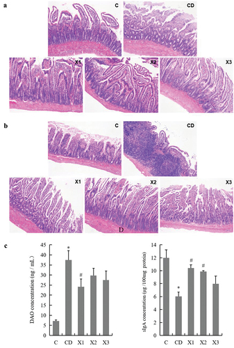
Figure 4. Xylitol ameliorated the small intestinal inflammation during the pathological process in STZ-induced diabetic rats. (A) Representative images of CD68+ cell infiltration (original magnifications, ×200). Formalin – fixed paraffin – embedded 5 μm cross-sections were stained with CD68+ primary antibody and analyzed for macrophage inflammatory infiltration. The concentration of inflammatory markers TNF-α (B) and IL-6 (C) in serum were evaluated by ELISA. The concentration of indicators of inflammatory infiltration myeloperoxidase (MPO) in serum and eosinophil peroxidase (EPX) in ileal tissue were detected using ELISA. Results are given as means ± SEM. Differences between groups were determined by ANOVA followed by Duncan’s test. * p < .05 vs control group; # p < .05 vs diabetic control group. C: control group; CD: diabetic control group; X1: 1.25 g/kg·bw xylitol; X2: 2.5 g/kg·bw xylitol; X3: 5 g/kg·bw xylitol.
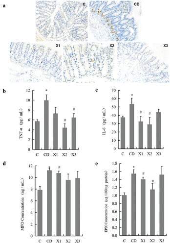
Figure 5. Xylitol preserves the mucosal barrier by improving the secretion of intestinal mucus. (A) and (B) are representative AB-PAS stained histological sections from the jejunum and ileum (original magnifications, ×100). C: control group; CD: diabetic control group; X1: 1.25 g/kg·bw xylitol; X2: 2.5 g/kg·bw xylitol; X3: 5 g/kg·bw xylitol.
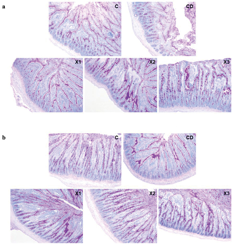
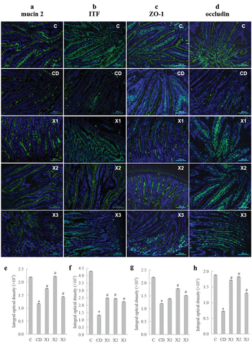
Figure 6. Xylitol preserves the mucosal barrier by improving the secretion of mucin and ameliorating the expression and distribution of tight junction proteins. Immunostaining of Carnoy-fixed tissue sections for (A) mucin-2 (anti-mucin-2 and FITC-anti-rabbit IgG, green; DAPI, blue) and (B) intestinal trefoil factor (ITF) (anti-intestinal trefoil factor and FITC-anti-rabbit IgG, green; DAPI, blue). (C) Immunostaining of jejunum tissue sections for ZO-1 (anti-ZO-1 and FITC-anti-rabbit IgG, green; DAPI, blue). (D) Immunostaining of jejunum tissue sections for occludin (anti-occludin and FITC-anti-rabbit IgG, green; DAPI, blue). (E) Integral optical density value of immunostaining of jejunum tissue sections for mucin-2, ITF(F), ZO-1(G), and occludin(H). Results are given as means ± SEM. Differences between groups were determined by ANOVA followed by Duncan’s test. * p < .05 vs control group; # p < .05 vs diabetic control group. C: control group; CD: diabetic control group; X1: 1.25 g/kg·bw xylitol; X2: 2.5 g/kg·bw xylitol; X3: 5 g/kg·bw xylitol.