
Figures & data
Table 1. Commercially available imaging systems
Table 2. Properties of selected fluorescent reporter proteins
Table 3. Imaging systems utilizing bioluminescent bacteria
Table 4. Comparison of Lux, GFP and mCherry
Figure 1. Lactobacillus plantarum 423 dam::cat-mCherry cells expressing the red fluorescent mCherry gene in the GIT of mice ex vivo. The color bar indicates the relative signal intensity measured as photons/sec/cm-2/sr. Image modified from van Zyl.Citation52
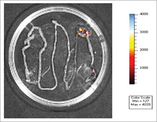
Figure 2. Arrangement of the luxCDABE open reading frames in Photobacterium spp, Vibrio spp. and Photorhabdus spp The luxI and luxR genes are adjacent to one another and luxI is the first of the 7 genes in the operon. luxI and luxR are required for activation of the luminescence genes.
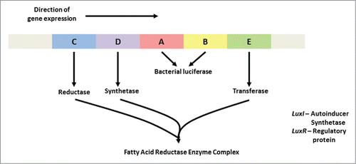
Figure 3. Monitoring of the colonization of Listeria monocytogenes EGDe in the intestinal tract of mice. Strain EGDe contains the luxABCDE operon of Photorhabdus luminescence. The bioluminescence signal was measured transcutaneously 30 min after infection, using an IVIS system (Caliper Life Sciences). The image was acquired using an integration time of 1 min. The color bar indicates the relative signal intensity measured as photons/sec/cm-2/sr. Image modified from van Zyl.Citation52
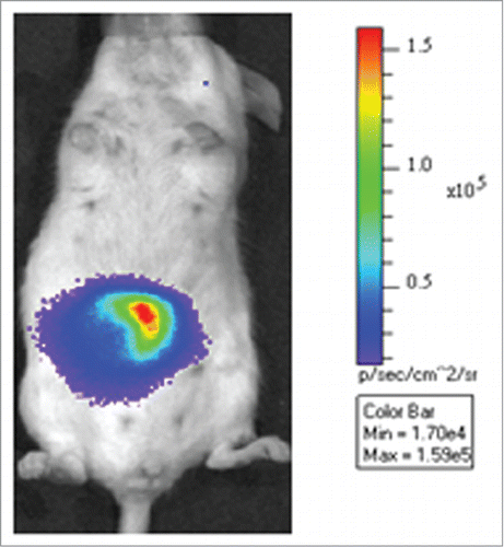
Table 5. Factors that can affect the sensitivity of imaging systems Citation27