
Figures & data
Figure 1. Subdoligranulum is associated with Akkermansia muciniphila and correlates positively with a healthy metabolic status
(A) Subset of the co-abundance network inferred using the SCS method related to the Akkermansia and Suddoligranulum genera. Nodes indicate metagenomic species and the two target genera, while edges indicate the inferred associations. Direction of the edges displays causal inference while the red and blue colors indicate respectively positive and negative associations. The colors of the vertices depict the different phylogenetic annotations at the family level – black being the two targets and white unknown phylogenetic annotations. (B) Heatmap of Spearman correlations between metagenomic species and clinical variables. ‘+’ indicates significant associations resisting adjustment for multiple testing while ‘*,’ associations which do not resist. N = 108.
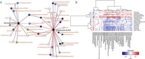
Figure 2. Impact of Subdoligranulum variabile supplementation on the prevention of diet-induced obesity in mice
(A) Study design: Mice were fed a high-fat diet (HFD) and gavaged with 1.5 × 10⁹ cultivable cells (cc) of freshly prepared life bacteria, 5 days a week for 8 weeks (HFD-Subdo group, n = 10, blue). This group was compared to mice that were gavaged with vehicle and were fed either a control diet (Control group, n = 11, green) or a high-fat diet (HFD group, n = 10, black). (B) Body weight (g) (C) lean mass (g) (D) and fat mass (g) evolution during the 8 weeks follow-up. (E) Cumulative food intake in kcal during the 8 weeks of treatment. (F) Weight in mg of different fat depots at the end of the experiment. SAT: subcutaneous (inguinal), EAT: epididymal, VAT: visceral (mesenteric), BAT: brown adipose tissue. (G) Weight in mg of different types of muscles. (H) qPCR quantification of Subdoligranulum at the end of a 5-day period of gavage. Data were analyzed using one-way ANOVA followed by a Tukey post hoc test for E-H or using 2-way repeated measures ANOVA for B-C. ‘*’ ‘**’ and ‘***’ indicate a significant difference versus HFD (P < .05, P < .01, P < .001, respectively).
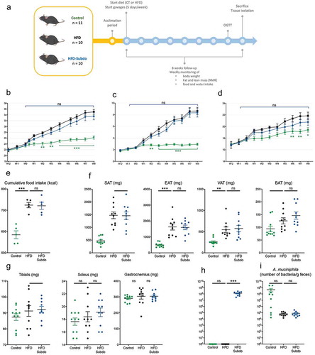
Figure 3. Subdoligranulum variabile supplementation does not impact on glucose metabolism
(A) Plasma glucose profile measured during an oral glucose tolerance test (OGTT). (B) Plasma insulin measured 30 min before and 15 min after glucose administration during the OGTT. (C) Mean area under the curve (AUC) of glucose measured during (OGTT). (D) Mean area under the curve of plasma insulin measured 30 min before and 15 min after glucose administration. (E) Insulin resistance index, calculated multiplying the area under the curve of both blood glucose (−30 to 120 min) and plasma insulin (−30 and 15 min) obtained following the oral glucose tolerance test. Data are represented as means ± SEM. Data were analyzed using 2-way repeated measures ANOVA for A or one-way ANOVA followed by a Tukey post hoc test for B-E.‘*’ ‘**’ and ‘***’ indicate a significant difference versus HFD (P < .05, P < .01, P < .001, respectively).
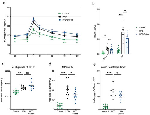
Figure 4. Subdoligranulum variabile supplementation does not impact on lipid metabolism
Effect of Subdoligranulum variabile administration on (A) liver weight (g), (B) liver lipid content (%), (C) liver triglycerides (mmol/mg), (D) liver cholesterol, (mmol/mg) (E) plasma triglycerides (mg/dl) and (F) plasma cholesterol (mg/dl). Data are represented as means ± SEM. Data were analyzed using one-way ANOVA followed by a Tukey post hoc test. ‘*’ indicates a significant difference versus HFD (P < .05).
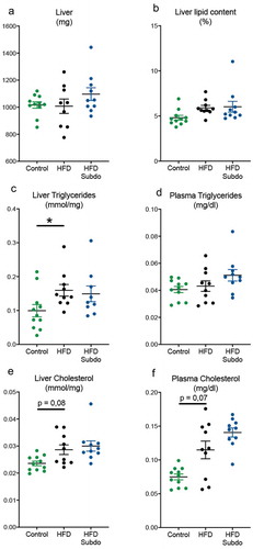
Figure 5. Subdoligranulum variabile supplementation does not impact on short-chain fatty acids or gut barrier
Effect of Subdoligranulum variabile administration on (A) short-chain fatty acids (nmol/g of dry cecal content) and (B) relative mRNA expression of key markers of gut barrier integrity. (ZO-1: Zonula occludens-1). Data are represented as means ± SEM. Data were analyzed using one-way ANOVA followed by a Tukey post hoc test.
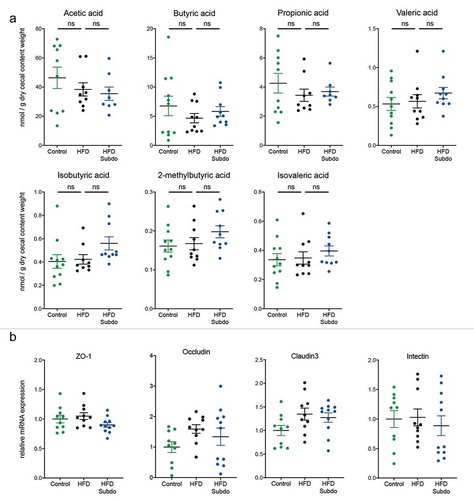
Table 1. Primers used for the qPCR
Supplemental material