987
Views
17
CrossRef citations to date
0
Altmetric
Articles

Antiproliferative properties of Saussurea lappa Clarke root extract in SH-SY5Y neuroblastoma cells via intrinsic apoptotic pathway

, &
Pages 119-126 | Received 11 Nov 2014, Accepted 24 Dec 2014, Published online: 11 Feb 2015

Figures & data

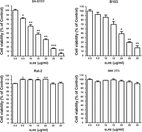
Figure 1. Antiproliferative and cytotoxic effects of SLRE in different cell lines. SH-SY5Y, B103, Rat-2, and NIH 3T3 cells were cultured in 96-well plates and treated with various concentrations of SLRE. After treatment for 24 h, CCK-8 (10 μl, Dojindo Lab) was added to each well of the plates and incubated for 3 h. A 96-well microtiter plate reader (Molecular Devices) was used to determine the absorbance at 450 nm for cell viability. Each point is the mean ± SEM of five samples. Data were composed of the means from three independent experiments in which the activity in the absence of SLRE versus in the presence of SLRE was found to be significantly different (n = 3, *p < 0.05, **p < 0.01, ***p < 0.001).
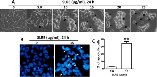
Figure 2. Morphological examination of SH-SY5Y cells treated with SLRE. (A) Cells were grown in 24-well culture dishes to near confluence (80%) and then treated with 0–25 μg/ml of SLRE for 24 h, after which morphology was observed by Bright-Field Microscopy. Arrows indicate cells with apoptotic morphology. (B) Apoptotic nuclear morphology was visualized by DAPI staining (arrow shows fragmented DNA). (C) The percentages of DAPI-stained cells were counted in three independent random areas. Results are means ± SE, and representatives of three independent experiments are shown (n = 3, **p < 0.01).
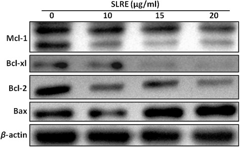
Figure 3. Regulation of apoptosis-related proteins in the Bcl-2 family by SLRE treatment in SH-SY5Y cells. Whole cell lysates were subjected to 15% SDS-PAGE and the levels of Mcl-1, Bcl-xl, Bcl-2, and Bax were detected by western blotting, as described in materials and methods. β-actin was used as a loading control.
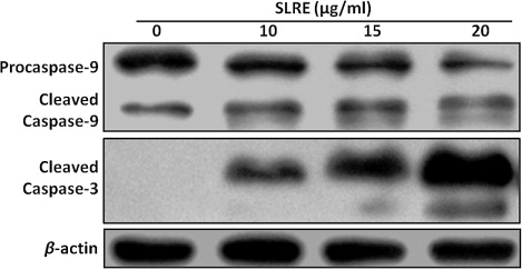
Figure 4. SLRE altered the expression of apoptosis-related proteins caspases-3 and -9 in SH-SY5Y cells. Cells were cultured in 60-mm culture dishes and treated with 0–20 µg/ml of SLRE for 24 h. Whole cell lysates were subjected to 15% SDS-PAGE and the levels of caspases-9 and -3 proteins were detected by western blotting.
Figure 5. Immunofluorescence analysis of cleaved caspase-3 in SLRE-induced apoptotic SH-SY5Y cells. Red fluorescence (Alexa Fluor 568 goat anti-rabbit) indicates cleaved caspase-3 expression, whereas the nucleus is stained blue (DAPI), as described in detail in “Materials and methods” section.
Figure 6. Effect of SLRE on AKT/GSK-3 signaling pathway in SH-SY5Y cells. Cells lysates were subjected to 10% SDS-PAGE and the levels of p-AKTSer473, AKT, p-GSK-3αSer21, p-GSK-3βSer9, p-GSK-3αtyr279/βtyr216, and GSK-3β proteins were detected by western blotting.

Reprints and Corporate Permissions

Please note: Selecting permissions does not provide access to the full text of the article, please see our help page How do I view content?

To request a reprint or corporate permissions for this article, please click on the relevant link below:

Academic Permissions

Please note: Selecting permissions does not provide access to the full text of the article, please see our help page How do I view content?

Obtain permissions instantly via Rightslink by clicking on the button below:

If you are unable to obtain permissions via Rightslink, please complete and submit this Permissions form. For more information, please visit our Permissions help page.