Figures & data
Figure 1. In vitro antioxidant activities of melittin and melittin-derived peptides. (A) Location of peptides in melittin protein. (B) ABTS radical scavenging activity of resveratrol (Res.), melittin, and melittin-derived peptides (P1–P4). Results are expressed as means ± SD (n = 3). *p < 0.05, ***p < 0.001.
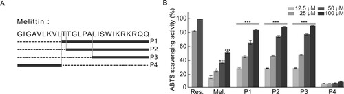
Figure 2. Comparison of cytotoxicity between melittin and melittin-derived peptides. (A-B) Cells were treated with specified concentrations of melittin and melittin-derived peptides for 24 h. Cell viability was determined using the MTS assay. Results are expressed as mean ± SD (n = 3). ***p < 0.001.
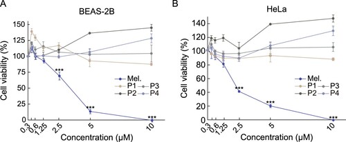
Figure 3. Comparison of anti-inflammatory activity between melittin and melittin-derived peptides. RAW 264.7 cells were pretreated with 1.25 μM melittin and melittin-derived peptides for 24 h and then incubated with LPS (1 μg/mL) for 6 h. qRT-PCR was performed to analyze the mRNA expression of the pro-inflammatory cytokines TNF-α (A), IL-6 (B), and IL-1β (C) Results are expressed as means ± SD (n = 3). (D-E) RAW 264.7 cells were pretreated with 1.25 μM melittin and indicated concentrations of peptide P1 and P2 for 24 h and then stimulated with LPS (1 μg/mL) for 1 h. The cell extracts were subjected to immunoblot analysis with anti-IκBα and anti-phospho-IκBα antibodies. Bar graphs show the relative expression of phospho-IκBα normalized with GAPDH. Results are expressed as means ± SD (n = 3). ***p < 0.001, **p < 0.01.
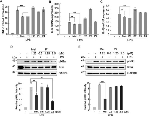
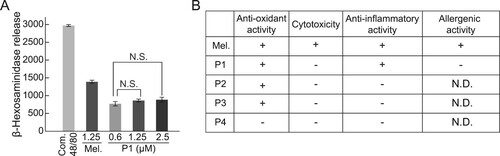
Figure 4. Comparison between degranulation activity of melittin and melittin-derived P1. (A) RBL-2H3 cells were treated with 1 mg/mL compound 48/80, 1.25 μM melittin, or indicated concentrations of P1 for 1 h. Compound 48/80 was used as a positive control for degranulation. Release of β-hexosaminidase was measured using fluorometric analysis. Results are expressed as mean ± SD (n = 3). N.S., no statistical significance. (B) Summary of activities of melittin and melittin-derived peptides. +, active. -, inactive. N.D., not determined.