Figures & data
Table 1. Clinico-pathological characteristics of the participants
Table 2. Table of the HER2 IHC and qRT-PCR
Figure 1. IHC staining images with (0, 1+, 2+, 3+) different scoring, were the 0, 1+ considered to be negative and 3+ considered to be positive
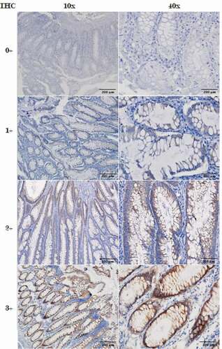
Figure 2. ERBB2 mRNA expression. Examples of real time PCR with SYBER Green. This was carried out in triplicate using RNA samples extracted from tumour cells microdissection from paraffin-embedded tissue sections
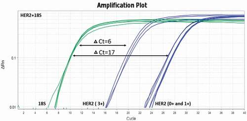
Figure 3. (a) Correlation of IHC group to HER2 protein expression and (b) correlation of HER2 mRNA expression between CRC study groups delta Ct mean to HER2 mRNA expression. * p < 0.05
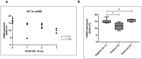
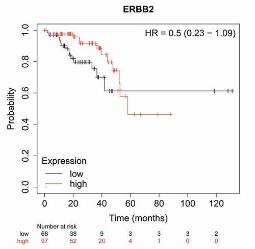
Figure 4. In large cohort of 165 patients, high HER2 expression show a trend, after 40 months, towards poor overall survival in rectum adenocarcinoma