Figures & data
Figure 1. The topography of the surface of the fixation plates
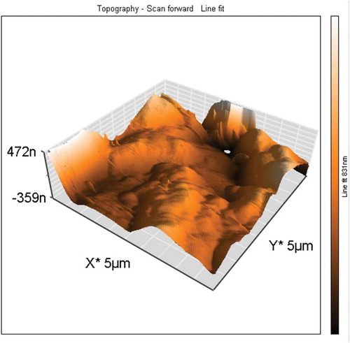
Table 1. The collective representation of the results of chemical composition microstructure analysis
Table 2. General printing parameters
Figure 2. Mean values and standard deviation of the assessment of metabolic activity in biofilms using TTC assay. This test demonstrated that after 48 h of incubation, all tested microorganisms present on the surface of the fixation plates showed metabolic activity in biofilms, but in a differentiated manner ()
Table 3. The mean values of surface roughness parameters, measured for three samples at five different places
Table 4. Average values of the number of cells recovered from the biofilm formed on the surface of the fixation plate model
Figure 3. (a), (b). Scanning electron microscopic image showing massive colonization by S. mutans.
Figure 4. (a), (b). Scanning electron microscopic image showing biofilm of S. epidermidis.
Figure 5. (a), (b). Scanning electron microscopic image showing biofilm of S. aureus.
Figure 6. (a), (b). Scanning electron microscopic image showing biofilm of L. rhamnosus.
Figure 7. (a), (b). Scanning electron microscopic image showing biofilm of C. albicans.