
Figures & data
Table 1. Patients’ characteristics.
Table 2. Agreement of the expert’s MRI scan readings (n = 30) with the respective MRI reports on file.
Figure 1. (a) Baseline diagnostic MRI of active-stage Charcot foot grade 0, four weeks after symptom onset. Sagittal STIR sequence showing EESC of tarsal bones (bright appearance), and soft tissue. (b) Same foot as in (a). First follow-up MRI after 6 weeks of unloading and immobilizing. Merely unchanged EESC, as compared to (a). (c) Second follow-up MRI after 11 weeks of treatment. Regression of bone and soft EESC (as compared to (a) and (b)). Unprotected normal weight-bearing was resumed immediately, without weaning. (d) Follow-up MRI after 21 weeks of unprotected re-loading. Relapse of bone EESC, now with tarsal fractures, soft-tissue edema, and collapse of the longitudinal arch, consistent with active-stage Charcot foot grade 1.
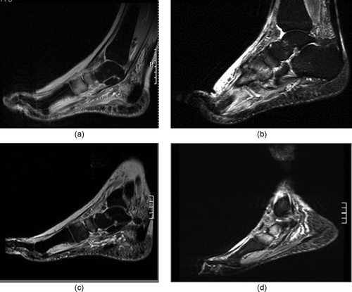
Table 3. Time intervals between sequential FUS.
Table 4. Cases and follow-up studies with regression of bone marrow EESC.