
Figures & data
Table 1. Extra-neural glioblastoma cases.
Figure 2. Patient's residual/recurrent glioblastoma patholog. (A) Histologic section of the residual/recurrent glioblastoma with giant cell features (black arrows), Hematoxylin and Eosin, scale bar 50 microns. (B) Whole genome copy number profile of residual/recurrent glioblastoma showing gains of chromosomes 7, 19 and 20, losses of chromosomes 10, 13, 18, 21 and 22 and amplification of EGFR on chromosome 7 (black arrow). (C) Histologic section of the lung mass demonstrating predominantly spindle cell neoplasm with areas of necrosis (black arrow) morphologically consistent with residual/recurrent glioblastoma with sarcomatous features. Hematoxylin and Eosin, scale bar 100 microns. (D) Histologic section of the lung mass demonstrating predominantly spindle cell neoplasm consistent with residual/recurrent glioblastoma with sarcomatous features, scattered multinucleated giant cells (black arrow) are present similar to the patient's intracranial tumor. Hematoxylin and Eosin, scale bar 50 microns. (E) Immunohistochemical stain for GFAP performed on lung mass showed scattered positive cells (black arrow), confirming the glial nature of the neoplasm. Scale bar 50 microns. (F) Whole genome copy number profile of lung mass showing gains of chromosomes 1p, 7 and 20, losses of chromosomes 4, 10, 13, 16, 18, 21 and 22 and amplifications of PDGFRA and CDK4 on chromosomes 4 and 12, respectively (black arrows).
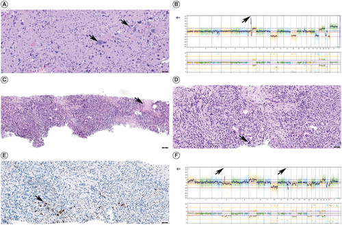
Figure 3. Patient's metastatic glioblastoma to the lungs. (A) Coronal computed tomography (CT) chest shows multiple enhancing pulmonary metastases. (B) Coronal CT chest shows further progression of pulmonary metastases and new hepatic metastases.
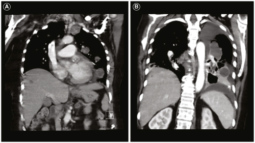
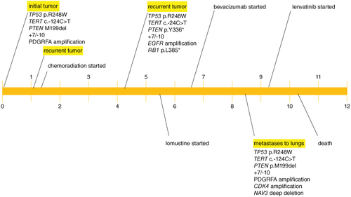
Figure 4. Patient disease course (timeline in months).
Data availability statement
The datasets from this study are available from the corresponding author upon reasonable request.