Figures & data
Figure 1. Preoperative epithelial thickness map of the case (X) in both eyes. The associated table divides the measurement to central 3mm (C) and paracentral nasal (N), temporal (T), inferior (I), and superior (S) 3–6mm annulus. Both eyes show almost the same epithelial thickness.
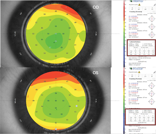
Figure 2. 1-week postoperative epithelial thickness map in both eyes for the same case. The associated table divides the measurement to central 3mm (C) and paracentral nasal (N), temporal (T), inferior (I), and superior (S) 3–6mm annulus. mPRK-treated eye shows increased central and paracentral epithelial thickness than tPRK-treated eye.
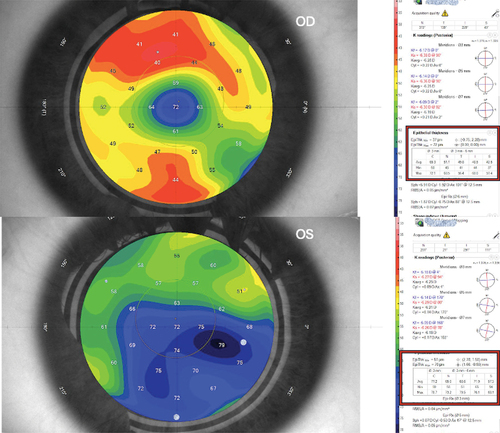
Figure 3. 6-month follow-up epithelial thickness map of the same case in both eyes. The associated table divides the measurement to central 3mm (C) and paracentral nasal (N), temporal (T), inferior (I), and superior (S) 3–6mm annulus. mPRK-treated eye shows slightly increased epithelial thickness than tPRK-treated eye.
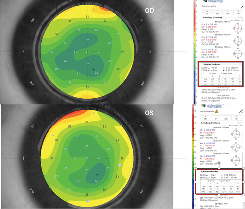
Table 1. Comparison between tPRK and mPRK according to spherical equivalent.
Table 2. Comparison between tPRK and mPRK according to Central epithelial thickness and mean Paracentral epithelial thickness.
Figure 4. Comparison between tPRK and PRK according to Central epithelial thickness.
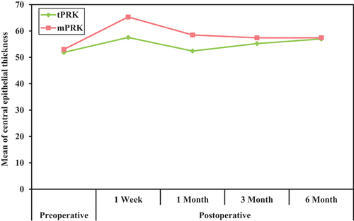
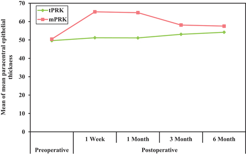
Figure 5. Comparison between tPRK and mPRK according to paracentral epithelial thickness.