Figures & data
Figure 1. Iron starvation leads to ROS accumulation and mitochondrial membrane damage in R. oryzae FTR1::RNAi mutant (inhibition of FTR1 expression) compared with control (RNAi empty). DHR-123 and Rh123 were used to measure ROS and mitochondrial membrane damage, respectively, using fluorescence microscopy and fluorescence spectrophotometry. Fluorescent images of R. oryzae FTR1::RNAi and control strain grown for 96 h in iron-deprived condition, stained with DHR-123 (A) and Rh-123 (C). Relative fluorescence of R. oryzae FTR1::RNAi and control strains stained with DHR-123 (B) and Rh-123 (D) grown in iron-deprived and iron-rich conditions. DIC, differential interference contrast. *P <0.05; ***P<0.0001 (compared with control strain).
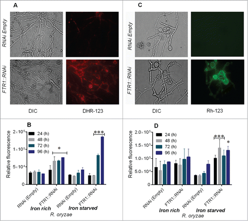
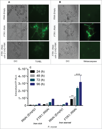
Figure 2. R. oryzae germlings deprived of iron display typical morphological changes associated with apoptosis, including nuclear degradation (by TUNEL assay), caspase-like activity (by CaspACE FITC-VAD-FMK), and extracellular ATP release (by CellTiter-Glo luminescent assay) in R. oryzae FTR1::RNAi and control strains grown for 24–96 h. Fluorescent images of R. oryzae FTR1::RNAi and control strains grown for 96 h in iron-deprived condition, stained with TUNEL (A), CaspACE FITC-VAD-FMK (B). Extracellular ATP release is higher in R. oryzae FTR1::RNAi mutant than control strain grown in iron-starved and iron-rich conditions, respectively (C). DIC, differential interference contrast). **p <0.001; ***p<0.0001 (compared with control).