Figures & data
Table 1. In vitro antimicrobial activity of CAS and MICA against C. albicans and C. parapsilosis strains biofilms
Figure 1. Fungicidal action of MICA against C. albicans (A–C) and C. parapsilosis (D–F) biofilms as shown by the morbidity stain DiBAC. (A) Fluorescence images of C. albicans stained with DiBAC. Relative fluorescence of DiBAC staining of CAS-S C. albicans (isolate 2) (B) and CAS-NS C. albicans (isolate 6) (C) treated with MICA. (D) Fluorescence images of C. parapsilosis stained with DiBAC. Relative fluorescence of DiBAC staining of CAS-S C. parapsilosis (isolate 7) (E) and CAS-NS C. parapsilosis (isolate 12) (F) treated with MICA. DIC: differential interference contrast; Ca: C. albicans; Cp: C. parapsilosis; C: untreated control. *P < 0.05; **P < 0.001; ***P < 0.0001 (compared with untreated control). The experiments were performed in triplicate and repeated 3 times.
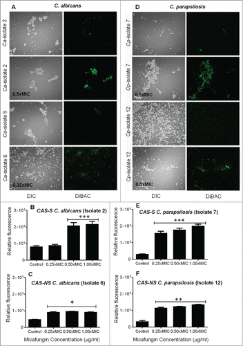
Figure 2. Extracellular β-1,3 glucan concentrations from C. albicans (A) and C. parapsilosis (B) planktonic and sessile cells. Each bar represents the mean and standard deviation from 2 replicates. *P < 0.05; **P < 0.001; ***P < 0.0001 (compared with untreated control).
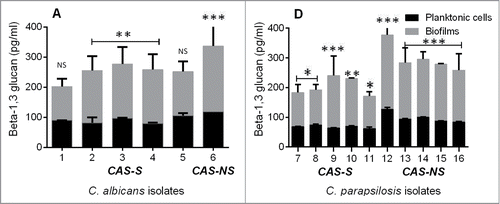
Figure 3. Intracellular ROS accumulation detected using fluorescence microscopy and fluorescence spectrophotometry in C. albicans (CAS-S isolate 2 and CAS-NS isolate 6) and C. parapsilosis (CAS-S isolate 7 and CAS-NS isolate 12) cells treated with MICA. Fluorescence images and relative fluorescence of CAS-S and CAS-NS strains of C. albicans (A, B) stained with DHR-123. Fluorescence images and relative fluorescence of CAS-S and CAS-NS strains of C. parapsilosis (C, D) stained with DHR-123. DIC: differential interference contrast; Ca: C. albicans; Cp: C. parapsilosis; C: untreated control. *P < 0.05; ***P < 0.0001 (compared with untreated controls).
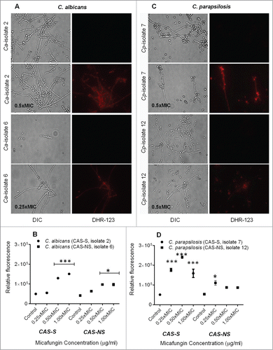
Figure 4. Measurement of depolarization of mitochondrial membrane potential by Rh-123 in C. albicans and C. parapsilosis sessile cells treated with MICA. Fluorescence images and relative fluorescence of CAS-S and CAS-NS strains of C. albicans (A, B) stained with Rh-123. Fluorescence images and relative fluorescence of CAS-S and CAS-NS strains of C. parapsilosis (C, D) stained with Rh -123. DIC: differential interference contrast; Ca: C. albicans; Cp: C. parapsilosis; C: untreated control. *P < 0.05; **P < 0.001; ***P < 0.0001 (compared with untreated control).
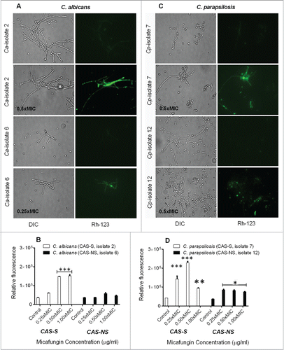
Figure 5. Detection of DNA fragmentation by TUNEL assay in CAS-S and CAS-NS C. albicans (isolates 2 and 6, respectively) and C. parapsilosis (isolates 7 and 12, respectively) sessile cells treated with MICA. Fluorescence images and relative fluorescence of CAS-S and CAS-NS strains of C. albicans (A, B) as detected by TUNEL assay. Fluorescence images and relative fluorescence of CAS-S and CAS-NS strains of C. parapsilosis (C, D) as detected by TUNEL. DIC: differential interference contrast; Ca: C. albicans; Cp: C. parapsilosis; C: untreated control. *P < 0.05; ***P < 0.0001 (compared with untreated control).
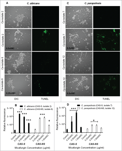
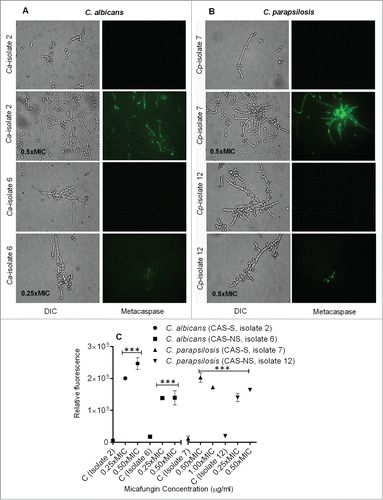
Figure 6. Caspase-like activity in CAS-S and CAS-NS C. albicans and C. parapsilosis strains treated with MICA. Fluorescence images of CAS-S and CAS-NS strains of C. albicans (A) and C. parapsilosis (B) treated with MICA and stained with CaspACE FITC-VAD-FMK. (C) Relative fluorescence of CAS-S and CAS-NS C. albicans and C. parapsilosis sessile cells stained with CaspACE FITC-VAD-FMK. DIC: differential interference contrast; Ca: C. albicans; Cp: C. parapsilosis; C: untreated control. ***P < 0.0001 (compared with untreated control).