Figures & data
Table 1. Oligonucleotide Sequences
Figure 1. Gene expression of Toll like Receptors, cytokines and antimicrobial peptides in corneal ulcers from patients with C. pseudodiphtheriticum keratitis. (A) Representative corneal ulcer of patient with C. pseudodiphtheriticum keratitis and Gram staining of corneal ulcer material showing Gram-positive C. pseudodiphtheriticum. RNA was isolated from corneal ulcers, reverse transcribed and quantitative PCR was done. Data points represent individual patients infected with C. pseudodiphtheriticum, and the values are presented either as log of relative expression or fold change in relation to uninfected donor corneas. The expressions of Toll like Receptors (B), proinflammatory cytokines (C) and antimicrobial peptides are (D) are shown. The representative gel images of the amplified products of antimicrobial peptides are shown (E). (**) designates P < 0.005.
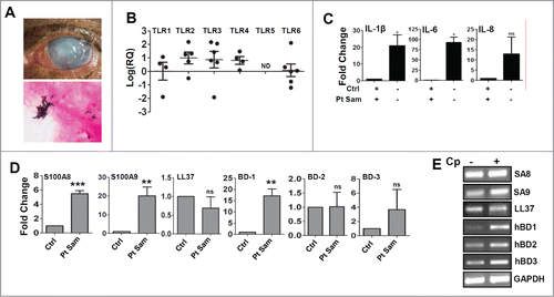
Figure 2. Expression and activation of Toll like Receptors by C. pseudodiphtheriticum in human corneal epithelial cells. Human corneal epithelial cells were infected with C. pseudodiphtheriticum for 4 hours, washed and RNA was isolated, reverse transcribed and quantitative PCR was done to determine the expression of Toll like Receptors in response to C. pseudodiphtheriticum (A); Western blot analysis of phosphorylation of IkBα in HCEC in response to C. pseudodiphtheriticum (B); p65 translocation from cytosol to nucleus in HCEC at 30 and 60 minutes postinfection with C. pseudodiphtheriticum was detected by immunocytochemistry (C). Activation of MAPK pathways in HCEC after incubation with C. pseudodiphtheriticum. At indicated times, cells were harvested and processed for Western blot analysis using antibodies to phospho-p38, phospho-ERK and phospho-JNK and β-actin (D).
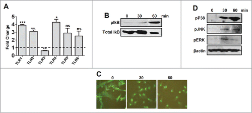
Figure 3. C. pseudodiphtheriticum causes expression of cytokines and activation of inflammasome. HCEC were infected with C. pseudodiphtheriticum for 4 hours, washed and RNA was isolated, reverse transcribed and expressions of cytokines (A) and inflammasomes (B) were determined by quantitative PCR. (*) designates P < 0.05.
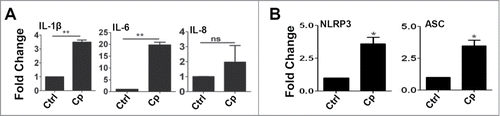
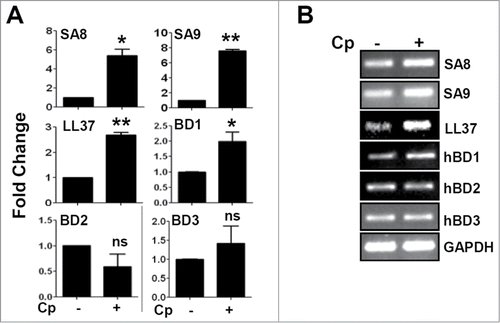
Figure 4. Up-regulation of antimicrobial peptide expression in response to C. pseudodiphtheriticum in human corneal epithelial cells. Immortalized human corneal epithelial cells were infected with C. pseudodiphtheriticum for 4 hours, washed and RNA was isolated, reverse transcribed and expressions of antimicrobial peptides were determined by quantitative PCR (A). The representative gel images of antimicrobial peptides are shown (B). (*) designates P < 0.05.