Figures & data
Figure 1. Cryptococcus neoformans (Cn) serotype A strains displayed higher survival percentage than serotype D strains after interactions with Acinetobacter baumannii (Ab). (A) Percentage survival of Cn serotype A and D strains after interaction with Ab. (B) Percentage survival of Ab after interaction with Cn strains. For A and B, bars are the averages of the results for 10 strains (each symbol represents an individual cryptococcal strain) per serotype, and error bars denote standard deviations (SDs). Asterisk denotes P-value significance (P < 0.05) calculated by Student's t-test. Each experiment was performed thrice with similar results obtained.
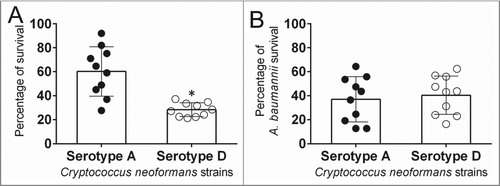
Figure 2. Cn serotype A strains form more metabolically active biofilms than serotype D strains after interactions with Ab. (A) Graphic representation of Cn and Ab biofilms interaction assay performed in this study. Fungal and bacterial biofilms were grown separately (Cn, 0.4 μm pore insert; Ab, bottom of a well) and co-incubated using a microtiter transwell system that permits chemotactic exchange through the supernatant. (B) Biofilm formation was determined by measuring the metabolic activity by XTT reduction assay. Bars are the averages of the results for 10 strains (each symbol represents an individual cryptococcal strain) per serotype, and error bars denote SDs. Asterisk denotes P-value significance (P < 0.05) calculated by Student's t-test. Each experiment was performed thrice with similar results obtained. (C) Confocal microscopy of Cn H99 and B3501 strain biofilms after interaction with Ab. Images of mature fungal biofilms showed metabolically active (red; FUN-1-stained) cells embedded in the polysaccharide extracellular material (green; stained with MAb 18B7-FITC-conjugated GAM IgG1). Images were obtained after 48 h co-incubation of the fungal cells to Ab. (D) The thickness and morphology of the cryptococcal biofilms can be observed in the Z-stack reconstruction. White arrows denote separation between Cn B3501strain biofilm aggregates. For C and D, the pictures were taken at a magnification of ×63. Bars, 20 µm. The results are representative of 2 distinct experiments.
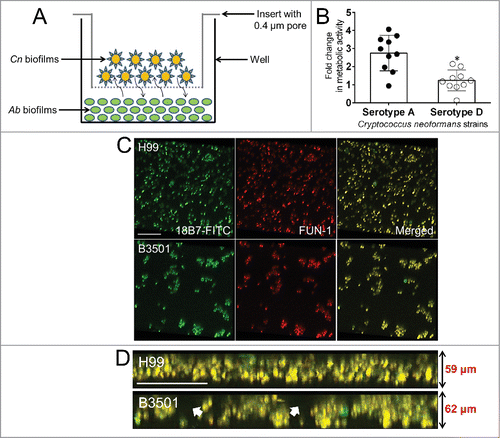
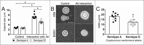
Figure 3. Impact of Ab interaction on Cn capsular size and GXM released. (A) Capsule size measurements of Cn serotype A and D strains were performed for yeasts grown in the absence and presence of Ab. Bars are the averages of the results for 25 cell measurements at each condition, and error bars denote SDs. (B) Representative India ink images displaying the effect of Ab interaction on the capsule size of Cn H99 (serotype A) and B3501 (serotype D). The pictures were taken using a ×100-power field. Scale bar, 2 µm. The experiments were performed thrice, and similar results were obtained. (C) GXM concentration in the supernatant of Cn serotype A and D strain cultures was determined by capture ELISA. Bars are the averages of the results for 10 strains per serotype, and error bars denote SDs. For A and C, asterisk denote P-value significance (P < 0.05) calculated by Student's t-test. Each experiment was performed thrice with similar results obtained.