Figures & data
Figure 1. Inactivation of HisB in A. fumigatus results in histidine auxotrophy (A) and growth inhibition of A. fumigatus by 3-AT is neutralized by histidine supplementation (B). Fungal strains were point-inoculated on Aspergillus minimal medium, complex medium and blood agar containing the indicated histidine concentrations and incubated at 37°C. Growth on minimal medium and blood agar was scored after 48 h, on CM after 24 h. For investigation of 3-AT-activity (B) minimal medium was used.
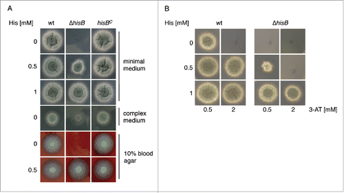
Figure 2. Histidine plays a crucial role in metal homeostasis of A. fumigatus. (A) Fungal strains were point-inoculated on minimal medium (MM) supplemented with the given concentrations of heavy metal salts, either with or without 1 mM externally added histidine. Photographs were taken after 48 h of incubation at 37°C. (B) Biomass production (dry weight) of wt and ΔhisB was quantified after growth for 24 h in liquid minimal medium with low contents of iron (-Fe), zinc (-zinc) or copper (-Cu) (see Material and Methods) as well as minimal medium containing additionally 5 mM FeSO4 (hFe), 4 mM ZnSO4 (hZn) or 1.5 mM CuSO4 (hCu). All cultivation conditions were performed with supplementation of either 0.5 mM or 5 mM histidine. Data represent the mean of 3 biological replicates ± standard deviation normalized to the biomass of the wt grown in minimal medium with the same histidine supplementation. The biomass of the wt in minimal medium was 0.78 ± 0.05 g at both 0.5 mM and 5 mM histidine concentrations supplemented. The decreased biomass production during metal starvation and excess underline the shortage and toxicity, respectively, of the metal concentrations.
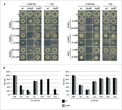
Figure 3. Histidine supplementation improves growth of the siderophore-deficient ΔsidA mutant (A) and decreases extra- and intracellular siderophore production (B). (A) Biomass production (dry weight) of wt and ΔsidA was quantified after growth for 21 h in liquid minimal medium under iron-replete (+Fe, 30 µM FeSO4) and iron depleted (-Fe) conditions with and without 5 mM histidine supplementation. Data represent the mean of 3 biological replicates ± standard deviation normalized to the biomass of the wt grown in +Fe conditions. The biomass of the wt in minimal medium +Fe was 0.63 g and 0.62 g with and without 5 mM histidine supplementation, respectively. (B) Intracellular (ferricrocin) and extracellular (TAFC) production of siderophores was quantified from the iron starvation conditions with and without 5 mM histidine supplementation (A) and normalized to the biomass and the wt grown without histidine supplementation.
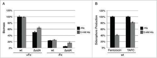
Figure 4. Hypoxia decreases histidine requirement of ΔhisB. Fungal strains were point-inoculated on minimal medium reflecting iron starvation (-Fe/BPS, containing 100 µM of the ferrous iron-specific chelator bathophenanthroline disulfonate), iron sufficiency (+Fe, 30 µM FeSO4), and iron excess (hFe, 5 mM FeSO4) and incubated at 37°C for 48 h in normoxic and hypoxic conditions, respectively. As a control for hypoxia, we included a mutant deficient in the transcription factor SrbA, ΔsrbA, which is essential for growth during hypoxic conditions unless supplemented with high iron concentrations. Citation50 Supplementation with 5 mM histidine resulted in wt-like growth of ΔhisB and did not affect the growth of ΔsrbA (data not shown).
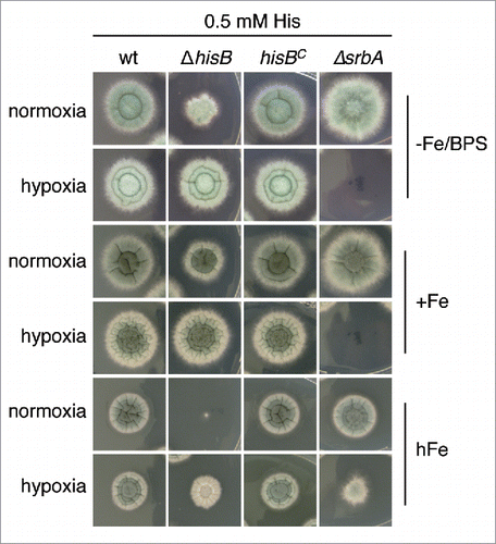
Figure 5. Histidine biosynthesis plays a crucial role in A. fumigatus virulence in G. mellonella. Larvae of the greater wax moth G. mellonella (n= 20 larvae per group) were infected with conidia of the respective strains and survival was monitored over a period of 6 d Control cohorts received either no injection (untreated) or were injected with the conidial solution buffer (IPS control). (A) HisB-deficiency (ΔhisB) attenuates virulence of A. fumigatus compared to the wt and hisBC strain (p <0.0001). (B) Co-injection of histidine increases virulence of ΔhisB (p<0.0018). (C) Co-injection of the histidine biosynthesis inhibitor 3-AT decreases virulence of the wt (p<0.0098).
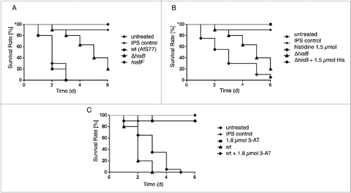
Figure 6. HisB-deficiency attenuates virulence of A. fumigatus in murine intranasal and intravenous infection models. (A) Survival curve for mice infected intranasally with 2 × 10⁵ conidia (p <0,001). (B) Survival curve for mice infected intravenously with 1 × 10⁵ conidia (p<0,001). (C) Histological analyses of mice infected intranasally with 2 × 10⁵ A. fumigatus conidia demonstrated that in contrast to ΔhisB, the wt and hisBC strains cause significantly increased cellular infiltration leading to major tissue damage. Murine lungs were embedded in Tissue-Tek O.C.T. (Sakura) and cryosections were co-stained with hemotoxylin-eosin and Grocott's Methenamine Silver according to standard protocols.
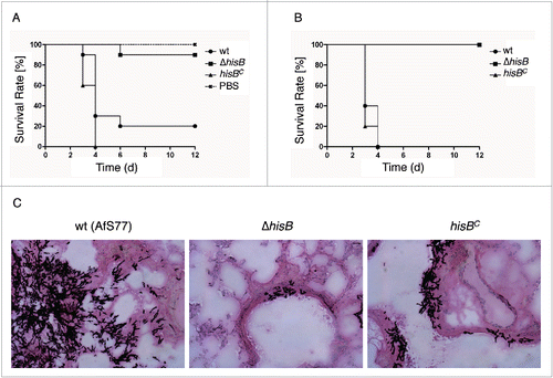
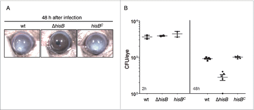
Figure 7. HisB-deficiency attenuates virulence of A. fumigatus in a murine model of fungal keratitis (A,B) and 3-AT treatment does not decrease the fungal burden (B). (A) 4 × 10⁴ A. fumigatus wt, ΔhisB or hisBC conidia were injected into the corneal stroma and corneal opacity and (B) CFU were measured at 48 h after infection (p <0.0).