Figures & data
Figure 1. MAbs inhibit SEK-induced proliferation of human immune cells. PBMCs from human whole blood were plated at 1 × 10⁵ cells per well and incubated in the presence of SEK for 96 hours at 37°C and 10% CO2. (A) SEK induces maximal proliferation of immune cells (up to 6-fold greater than controls) at 10ng/ml. Immune cell proliferation after treatment with 10ng/ml of SEK is inhibited in a dose-dependent manner by concurrent incubation with 1, 10, and 100ug/ml of (B) mAb-4G3, (C) mAb-5G2, and (D) mAb-9H2. Relative luminescence units (RLU); Treatment with media alone (RPMI); Murine antibody isotype control (IC); Student's t-test: * = p < 0.05; ** = p < 0.01.
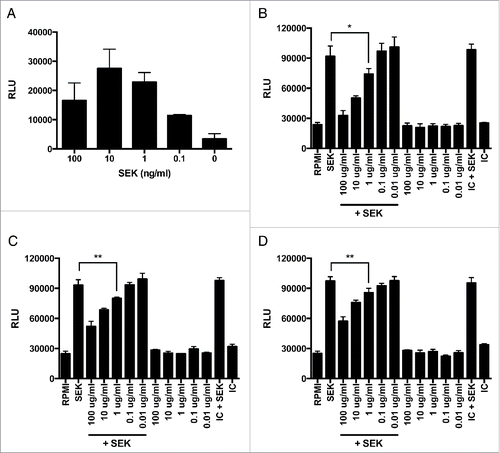
Figure 2. MAbs inhibit SEK-induced cytokine secretion by human immune cells. T cells from human whole blood were plated at 5 × 10⁴ cells per well and incubated in the presence of SEK and 1ug/ml of mAb(s) for 8 hours at 37°C and 10% CO2. Supernatants were subsequently collected and pro-inflammatory cytokines were measured by ELISA for human (A) IL-2 or (B) INF-γ. Treatment with media alone (RPMI); Murine antibody isotype control (IC). Student's t-test: * = p < 0.05; ** = p < 0.01; *** < 0.001.
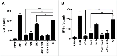
Figure 3. SEK binding competition between mAb combinations. A 96-well ELISA plate was coated with 25ng of SEK per well, blocked with 1% BSA, and then simultaneously treated with (A) the combination of mAb-4G3 (IgG2b) and mAb-5G2 (IgG1) or (B) the combination of mAb-9H2 (IgG1). MAb-4G3 and mAb-5G2 do no exhibit competition for epitope(s) on SEK. mAb-4G3 and mAb-9H2 exhibit competition for epitope(s) on SEK. All experiments were performed in duplicate. GAM = Goat anti-mouse, AP = Alkaline phosphatase.
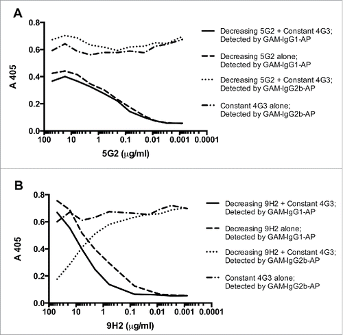
Table 1. Characterization of SEK monoclonal antibodies.
Table 2. The combination of non-competing mAbs protects mice from SEK-induced lethal shock.
Table 3. Competitive mAb combination does not protect from SEK lethal shock.
Figure 4. MAb combination is protective against sepsis by S. aureus USA300. (A) 6-8 week old mice female BALB/c mice were intravenously administered 1mg of mAb-4G3 or mAb-5G2 or both (1:1: ratio) or an equivalent volume of PBS control, followed within 1 hr by intravenous administration of 5.1 × 10⁶ CFUs of USA300 strain, B-155. Where indicated, vancomycin was administered once daily for 3 days starting 24 hrs before infection. Survival curves were analyzed by Log-rank (Mantel-Cox) test (GraphPad Prism 6 software). (B) Body weight was monitored daily during the course of 21-day experiment. Change in body weight is relative to weight of same group of mice on Day 0, prior to bacterial challenge. Mann-Whitney test (GraphPad Prism 6 software): * P ≤ 0.05, ** P ≤ 0.01.
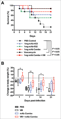
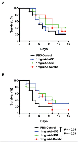
Figure 5. mAb booster is mildly protective from sepsis with SEB+ s. aureus. (A) Six to 8 week-old female BALB/c mice were intravenously administered 1mg of mAb-4G3 (IgG2b) or mAb-5G2 (IgG1) or both (1:1 ratio) or an equivalent volume of PBS control, followed within 1 hr by intravenous administration of 1.1 × 10⁸ CFUs of MRSA strain W-132. (B) Similarly to panel A, mice were intravenously administered 1.2 × 10⁸ CFUs of W-132, and received an additional intravenous administration of antibody or PBS control at 5 days post-infection. Survival curves were analyzed by Log-rank (Mantel-Cox) test (GraphPad Prism 6 software).