
Figures & data
Figure 1. Full-length and truncated B19-VP1u (tVP1u) proteins. (A) Schematic representation of full-length (1) and deletions of B19-VP1u. Eleven truncated B19-VP1u recombinant proteins correspond to amino acid sequences 21–227 (2), 31–227 (3), 51–227 (4), 61–227 (5), 71–227 (6), 82–227 (7), 91–227 (8), 101–227 (9), 111–227 (10), 121–227 (11) and 130–227 (12). Separation of (B) total lysate and (C) purified B19-VP1u proteins in a 12.5% SDS-PAGE. (D) Identification of purified recombinant B19-tVP1u proteins with anti-histidine monoclonal antibodies. Lane M indicates pre-stained protein marker; lane 1 indicates full-length B19-VP1u, and lanes 2 to 12 correspond to truncated deletions 2 to 12, respectively.
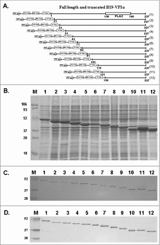
Table 1. Secreted phospholipase A2 (sPLA2) activity of recombinant full -length and N-terminal truncated B19-VP1 unique region (B19-tVP1u) proteins.
Figure 2. Reactivity of antibodies against cardiolipin and β2GPI to truncated B19-VP1u (B19-tVP1u) proteins. Full-length B19-VP1u and B19-tVP1u proteins were probed with antibodies against (A) cardiolipin and (B) β2GPI. Lane M indicates pre-stained protein marker; lane 1 indicates the full-length B19-VP1u, and lanes 2 to 12 correspond to the truncated deletions 2 to 12, respectively.
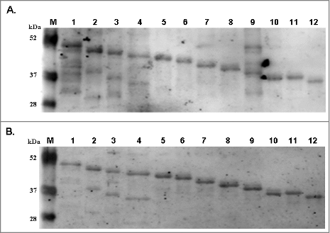
Figure 3. Binding activity of antibodies against B19-tVP1u with CL and β2GPI. IgG against B19-tVP1u was purified from BALB/c mice and reacted with (A) CL and (B) β2GPI. By ELISA analysis, normal absorbance of controls was 0.191 ± 0.03 (mean ± 2 SD) for CL and 0.137 ± 0.01 (mean ± 2 SD) for β2GPI. # indicates significance, P<0.05, relative to the controls (CTL). N = 4 mice per group.
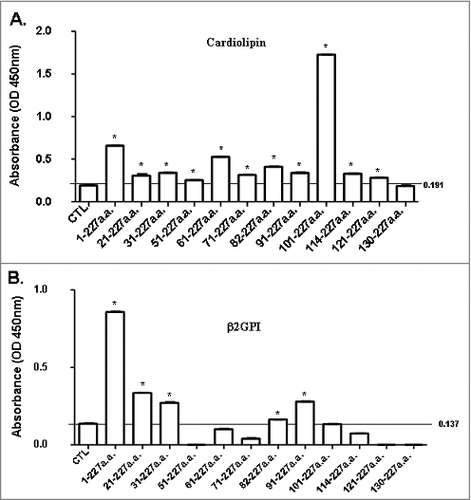
Table 2. Absorption of mouse anti-B19-VP1u or B19-tVP1u antibodies with purified B19-VP1u or B19-tVP1u proteins for binding of cardiolipin autoantigen.
Table 3. Absorption of mouse anti-B19-VP1u or B19-tVP1u antibodies with purified B19-VP1u or B19-tVP1u proteins forβ2GPI autoantigen.
Table 4. Mice infused with various B19-tVP1u antibodies.