Figures & data
Figure 1. The three steps of the T. cruzi invasion. The scheme summarizes the process of T. cruzi entry into the host cell. (1) Adhesion of trypomastigotes to the host cell surface. (2) Internalization of trypomastigotes produced by invagination of the plasma membrane. (3) Vacuole maturation proceeds after the fusion with lysosomes which initiates the differentiation of T. cruzi from trypomastigotes to amastigotes.

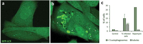
Figure 2. Autophagy is induced during T. cruzi infection. CHO cells overexpressing GFP-LC3 were infected with tissue culture trypomastigotes (TCT) of T. cruzi Y strain (MOI = 50) during 3 h. After fixation, cells were washed, mounted in mowiol and analyzed by confocal microscopy. (a) Control (non-infected) cells depicting the homogeneous distribution of GFP-LC3 in the soluble form. (b) Infected cell displaying numerous autophagic vacuoles and tubules (arrows) decorated with membrane-associated GFP-LC3. Note the recruitment of this protein in the membrane of the T. cruzi parasitophorous vacuole (asterisks) that were recognized by the typical form of trypomastigotes. (c) Percentage of cells containing more than 5 LC3-positive vesicles/cell and at least one tubule/cell at the indicated conditions. Rapamycin (50 ng/μl) were incubated for 2 h in control conditions. Scale bar: 5 μm. Data shown represent the mean ± SE from 3 independent experiments. *p < 0.05, ***p < 0.001 (Student´s t-test).