Figures & data
Table 1. Presence of replicating virus in different organs of the gravid mice.
Figure 1. Schematic diagram: Gestational (G) period, embryonic (E) age, infection timings, and organ collection schedules.
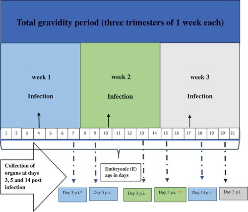
Figure 2. Viral RNA load in pancreas of dams infected during gravidity.
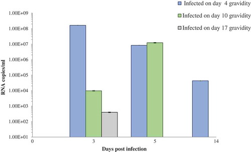
Figure 3. (a) Viral RNA load in pancreas of the challenged pups of CVB4-E2-infected and control dams. (b) Viral RNA load in pancreas of “individual” challenged pup showing positivity at day 12 postinfection of CVB4-E2-infected and control dams. (c) Viral RNA load in pancreas of “individual” challenged pups showing positivity at day 21 postinfection of CVB4-E2-infected and control dams.
*+/+ both mother and pup infected; −/+ mother mock-infected and pup-infected, P = pup; 1, 2, 3, 4, 5 number code of individual pup for that catergory. Days 4, 10, and 17 show the time of infection of the dam.
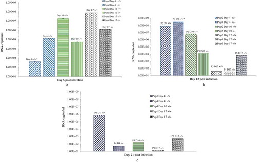
Table 2. Grades of destruction in the pancreas tissues of CVB4-E2 infected dams.
Figure 4. Histopathological changes in pancreas tissues of CVB4-E2 infected and control dams. (a) Absence of changes in the acinar and endocrine pancreas tissue (20×) and in the peripancreatic fat tissue (20×) (b) of a dam mock infected on day 4/E.4 sacrificed on day 3 p.i./E.7. (c) Focal infiltration in the peripancreatic fat tissue of virus-infected dam on day 10/E.10, sacrificed on day 3/E.13 p.i. (20×). (d) Pancreatitis in the acinar tissue showing inflammatory phase in the tissue of dam infected with the virus on day 4/E.4 sacrificed on day 3 p.i./E.7 (20×). (e) A stage of transition from acute to a subacute condition with reparative changes, lymphoplasmacytic inflammation with several incipient capillaries, and formation of inflammatory granulation in the defunct parenchymal pancreatic tissue. The lymphocytic infiltrates were also observed in the pancreatic islets of dams infected with the virus on day 10/E.10 (20×) and sacrificed on day 5 p.i./E.15.
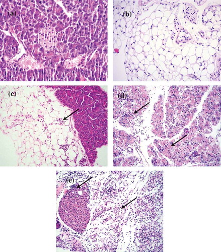
Table 3. Histopathology of pancreas tissues of challenged pups of dams infected in the first week of gestation (day 4/E.4).
Table 4. Histopathology of pancreas tissues of challenged pups of dams infected in the second week of gestation (Day 10/E.10).
Table 5. Histopathology of pancreas tissues of challenged pups of dams infected in the third week of gestation (day 17/E.17).
Figure 5. Histopathological changes in the pancreatic tissues of the virus challenged pups of CVB4-E2 infected and pups of mock control dams.
(a) Absence of changes in the acinar and endocrine pancreatic tissue (20×) dam infected on day 4/E.4 of gestation and virus challenged pup sacrificed on day 5. (b) Peripancreatic fat tissue (20×) of a mock-infected dam on day 4/E.4 of gestation and (−/−) pup mock challenged and sacrificed on day 5 p.i. (c) Mild and severe (d) forms of infiltration in the peripancreatic fat tissue of a virus challenged two different pups (−/+) of a dam mock-infected on day 4/E.4 of gravidity (20×), pups sacrificed on day 5 p.i. (20×). (e) Chronic pancreatitis and atrophy of the acinar tissue of a challenged pup (+/+) sacrificed on day 5 p.i. born to dam infected with virus on day 10/E.10 of gravidity, (f) chronic pancreatitis and atrophy of the acinar tissue and infiltration in the endocrine islet of a challenged pup (+/+) sacrificed on day 21 p.i. (20×) born to dam infected with the virus on day 10/E.10 (20×) in the tissue of pup sacrificed on day 21 p.i., (g) chronic pancreatitis in the acinar tissue of the virus challenged pups (+/+) of dam infected on day 17/E.17 of gestation day, pup sacrificed on day 12 p.i., absence of infiltration of the islets (20×), (h) chronic pancreatitis and atrophy in another challenged pup (+/+) sacrificed on day 12 p.i. (20×) of dams infected on day 17/E.17 of gestation day.
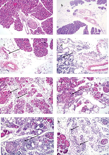
Figure 6. Localization of VP1 in the pancreatic tissue. (a) Absence of VP1 in the islet and exocrine tissue of a mock-infected pup of a dam mock infected on day 4/E.4 (−/−) (20×), pup sacrificed on day 5 p.i. (b) Presence of VP1 in the islet and exocrine tissue on day 3 p.i./E.20 of a dam infected with CVB4-E2 on day 17/E.17. (c) Presence of VP1 in the exocrine tissue but absence in the islet of virus challenged pup of a dam infected on day 10/E.10 (+/+), and of dam at day 17/E17. (d) Pups sacrificed on day 5 p.i. (20×). (e) Peripancreatic fat tissue showing VP1 positivity at day 5 p.i. in the pancreas of a virus challenged pup of a mock-infected dam (20×).
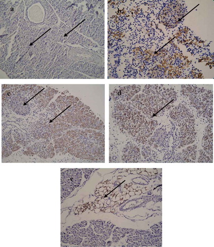
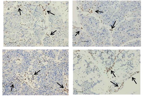
Figure 7. CD11b+ macrophage and LY6G+ neutrophils localization.
(a) CD11b+ macrophage and (c) LY6G+ neutrophils in abundance seen in the pancreas tissue infected with CVB4-E2 on day 4 (E.4), collected on day 3 p.i. from a dam; (b) CD11b+ macrophages observed in the pancreas tissue collected on day 5 p.i. of a challenged pup of dam infected with CVB4-E2 on day 4 (E.4) whereas (d) shows CD11b+ macrophages seen in the pancreas tissue collected on day 21 p.i. of a challenged pup of dam infected with CVB4-E2 on day 17 (E.17).