
Figures & data
Figure 1. S. argenteus and S. schweitzeri isolates are cytotoxic to human cancer cells. Broth from overnight cultures of S. argenteus RK308 (RK308), S. argenteus MSHR1132T (S. arg), S. schweitzeri FSA084T (S. schw), S. aureus 1800T (S. aur) and MRSA CCUG 35601 (MRSA) was added to HeLa and HT29 cells for 6 h. Un-inoculated broth was used as mock. (a) Cells were photographed through the microscope lens. (b) Viability of cells from (a) calculated from the MTT assay compared to mock, which was set to 100%. Asterisks above bars show significant differences as compared to S. aureus 1800T.
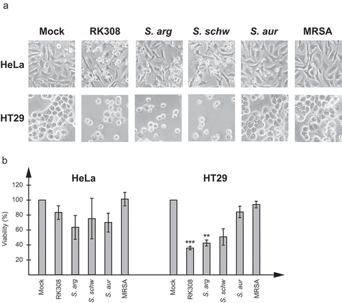
Figure 2. Identification of the cytolysin by MS analyses. (a) Coomassie brilliant blue stained SDS-PAGE of bacterial broth from S. argenteus RK308 (RK308), S. argenteus MSHR1132T (S. arg), S. schweitzeri FSA084T (S. schw), S. aureus 1800T (S. aur) and MRSA CCUG 35601 (MRSA) used in the cytotoxicity assay. Bands 1, 2 and 3 from the RK308 lane were excised from the gel for MS analyses. MWM, molecular weight marker. (b) Complete amino acid sequence of alpha-hemolysin, Hla, with peptides identified by MS shown in bold.
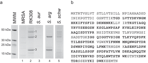
Figure 3. Neutralization of the cytotoxic effect by an anti-Hla antibody. Broth from overnight cultures of S. argenteus RK308 (RK308), S. argenteus MSHR1132T (S. arg), S. aureus 1800T (S. aur) and MRSA CCUG 35601 (MRSA) was added to HT29 cells for 6 h together with the anti-Hla antibody or PBS as indicated. Un-inoculated broth was used as mock. (a) Cells were photographed through the microscope lens. (b) Viability of cells from (a) calculated from the MTT assay compared to mock, which was set to 100%. Asterisks show significant differences between antibody- and PBS-treated cells.
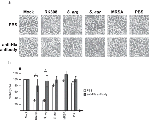
Figure 4. S. argenteus expresses high levels of alpha-hemolysin, hla. Expression analyses of hla and regulatory genes in S. argenteus RK308 (RK308), S. argenteus MSHR1132T (S. arg), S. schweitzeri FSA084T (S. schw), S. aureus 1800T (S. aur) and MRSA CCUG 35601 (MRSA). (a) Detection of the hla gene using PCR. NTC, no template control. (b) RNA levels of hla detected with RT-qPCR. RNA expression levels are normalized against 16SrRNA and expressed as fold compared to the S. aureus 1800T isolate. Asterisks above bars show significant differences as compared to S. aureus 1800T. (c) Hla protein levels in bacterial broth used in the cytotoxicity assay detected by western blot with an anti-Hla antibody. B, un-inoculated broth. Numbers below the blot show fold expression levels compared to S. aureus 1800T as quantified in Photoshop. (d) Schematic diagram showing the regulation of hla transcription and translation by regulatory factors. Adapted from [Citation25]. (E) RNA levels of regulatory genes sarA, saeR, and RNAIII detected with RT-qPCR. RNA expression levels are normalized against 16SrRNA and expressed as fold compared to the S. aureus 1800T isolate. Asterisks above bars show significant differences as compared to S. aureus 1800T.
![Figure 4. S. argenteus expresses high levels of alpha-hemolysin, hla. Expression analyses of hla and regulatory genes in S. argenteus RK308 (RK308), S. argenteus MSHR1132T (S. arg), S. schweitzeri FSA084T (S. schw), S. aureus 1800T (S. aur) and MRSA CCUG 35601 (MRSA). (a) Detection of the hla gene using PCR. NTC, no template control. (b) RNA levels of hla detected with RT-qPCR. RNA expression levels are normalized against 16SrRNA and expressed as fold compared to the S. aureus 1800T isolate. Asterisks above bars show significant differences as compared to S. aureus 1800T. (c) Hla protein levels in bacterial broth used in the cytotoxicity assay detected by western blot with an anti-Hla antibody. B, un-inoculated broth. Numbers below the blot show fold expression levels compared to S. aureus 1800T as quantified in Photoshop. (d) Schematic diagram showing the regulation of hla transcription and translation by regulatory factors. Adapted from [Citation25]. (E) RNA levels of regulatory genes sarA, saeR, and RNAIII detected with RT-qPCR. RNA expression levels are normalized against 16SrRNA and expressed as fold compared to the S. aureus 1800T isolate. Asterisks above bars show significant differences as compared to S. aureus 1800T.](/cms/asset/29164868-e265-4ab9-9629-6c188c749706/kvir_a_1620062_f0004_oc.jpg)