Figures & data
Figure 1. Enterovirus 71 Exit Cells Non-lytically In Vitro. (a) viral RNA was detected in culture supernatant after infected 1–9 h by qRT-PCR assay (mean±SD; three independent experiments). (b) infected cells incubated with SYTOXTM Blue Nucleic Acid Stain to monitor plasma membrane intactness. c, quantification of fluorescence intensity in ) (mean±SD; three independent experiments; *p ≤ 0.05). Statistical analysis were performed using unpaired two-tailed student’s t-test. D, single-step growth curve of RD cells infected with EV71 at MOI of 1. Supernatants were collected from infected cells at the indicated time points, and virus titers were calculated by standard TCID50 assay
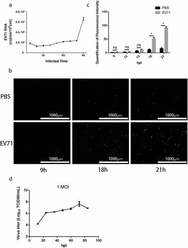
Figure 2. EV71 virions could be wrapped within exosomes in vitro. (a) buoyant density of EV71 particles released by RD cells in iodixanol gradients. Exosome-like vesicles wrapping EV71 particles (EXO-EV71) gathered in 1.10–1.12g/mL (fraction 7–8), non-enveloped EV71 particles gathered in 1.18–1.26g/mL (fraction 4–5). (b) TEM images of EXO-EV71 (a-c, fraction 7–8 in A) and non-enveloped EV71 (d, fraction 4–5 in A). c, distribution of the number of virion-like particles contained within individual exosomes. d, distribution of EXO-EV71 size measured by nanoparticle NTA. e, immunoblots of EV71 capsid proteins (VP1) and exosomes positive marker (CD63 and TSG101) and negative marker (CANX and Albumin) in lysate of EV71-infected cells, EXO-EV71 and EV71 particles. f, immunoblot analysis of CD63, TSG101 and VP1 in EXO-EV71 exposed to proteinase K with or without detergent (0.1% Triton X-100, used to ensure degradation of both surface and internal components)
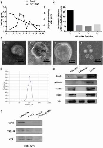
Figure 3. Exosomes Containing Viral RNA (EXO-EV71-RNA) Existed in Patients’ Plasma. (a) viral RNA could be detected in patients’ plasma and in the exosomes of the plasma. (b) viral RNA was positive in patients’ stools