Figures & data
Table 1. Sequences of primers, miRNAs, and siRNAs used in this study
Figure 1. CSFV infection down-regulates miR-140 expression
(a) The standard curve of for detecting CSFV RNA genome. (b) Quantification of CSFV viral genomic RNA in supernatant. The copy number of CSFV viral genomic RNA is calculated by using the standard curve in (a). Cells were seed in a 48-well plate and infected with CSFV (MOI = 3). Then, the supernatant was collected at 0, 6, 12, 24, 48, and 72 h post-infection. (c) Real-time qRT-PCR analysis of miR-140 expression (normalized to U6 expression). The SUVECs were infected with CSFV (MOI = 0.1) and were collected at 12, 24, 48, and 60 h post-infection. Results are expressed as mean ± SD of three independent experiments. p values were calculated using Student’s t-test. *P < 0.05, ***P < 0.001.
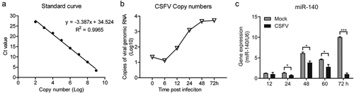
Figure 2. miR-140 mimics inhibits CSFV replication
(a and b) SUVEC were transfected with various concentrations of miR-140 mimics (30, 60, and 150 nM) or mimic control (MC) and infected with CSFV (MOI = 0.1). (a) Real-time qRT-PCR analysis of CSFV viral RNA (normalized to β-actin expression) at 48 h post-infection. (b) Quantification of CSFV viral genomic RNA in supernatant. The copy number of CSFV viral genomic RNA is calculated by using the standard curve in ()). (c and d) SUVEC were transfected with of miR-140 mimics (60 nM) or mimic control (MC) and infected with CSFV (MOI = 0.1). The sample was collected at 24, 36, 48, and 72 h post-infection. (c) Real-time qRT-PCR analysis of CSFV viral RNA (normalized to β-actin expression). (d) Quantification of CSFV viral genomic RNA in supernatant. The copy number of CSFV viral genomic RNA is calculated by using the standard curve in ()). Results are expressed as mean ± SD of three independent experiments. p values were calculated using Student’s t-test. *P < 0.05, **P < 0.01.
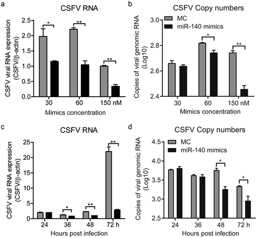
Figure 3. miR-140 inhibitor promotes CSFV replication
SUVECs were transfected with miR-140 inhibitors (200 nM) or inhibitor control (IC) and infected with CSFV (MOI = 0.1). (a) Real-time qRT-PCR analysis of CSFV viral RNA (normalized to β-actin expression) at 12, 24, 36, 48 h post-infection. (b) Quantification of CSFV viral genomic RNA in supernatant at 12, 24, 36, 48 h post-infection. The copy number of CSFV viral genomic RNA is calculated by using the standard curve in ()). Results are expressed as mean ± SD of three independent experiments. p values were calculated using Student’s t test. *P < 0.05, **P < 0.01, ***P < 0.001.
Figure 4. MiR-140 binds to the 3ʹ UTR of Rab25
(a) RNA22 V2 and RNAhybrid online software were used to verify the target gene of miR-140. Alignments showed that there were two complementary sequences between miR-140 sequences and Rab25-3ʹ UTR sequences. Underlined sites indicate miR-140 seed region and mutated sequences are shown. (b) Dual-luciferase assays. Wild-type (WT) (pmirGLO-Rab25-WT) or mutant (pmirGLO-Rab25-Mut) luciferase-reporter vector and miR-140 mimics were co-transfected into cells. Luciferase activity were determined at 48 h post-transfection by dual-luciferase assays. (c, d, and e) 60 nM miR-140 mimics or mimics control (MC) were co-transfected into cells. (c) Real-time qRT-PCR analysis of miR-140 expression (normalized to U6 expression) at 48 h post-infection. (d) Real-time qRT-PCR analysis of Rab25 expression (normalized to β-actin expression) at 48 h post-infection. (e) Immunoblot analysis of Rab25 protein levels at 48 h post-infection. The number means the intensity of Rab25 protein level (top) normalized to that of β-actin. (f, g, and h) 60 nM miR-140 inhibitor or inhibitor control (IC) were co-transfected into cells. (f) Real-time qRT-PCR analysis of miR-140 expression (normalized to U6 expression) at 48 h post-infection. (g) Real-time qRT-PCR analysis of Rab25 expression (normalized to β-actin expression) at 48 h post-infection. (h) Immunoblot analysis of Rab25 protein levels at 48 h post-infection. The number means the intensity of Rab25 protein level (top) normalized to that of β-actin. Results in (a, c, d, f, and g) are expressed as mean ± SD of three independent experiments. p values were calculated using Student’s t-test. Western blots were analyzed and quantified using the Image J software. *P < 0.05, **P < 0.01, ***P < 0.001.
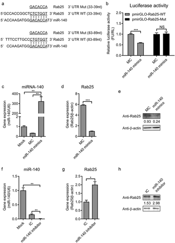
Figure 5. The expression of Rab25 is upregulated after CSFV infection in SUVECs
(a) Real-time qRT-PCR analysis of Rab25 expression (normalized to β-actin expression) at 12 h, 24 h, 36 h, and 48 h after CSFV infection in SUVEC. (b) Immunoblot analysis of Rab25 protein levels. The treatment is the same as (b). Results in (a) were expressed as mean ± SD of three independent experiments. p values were calculated using Student’s t-test. Western blots were analyzed and quantified using the Image J software. *P < 0.05.
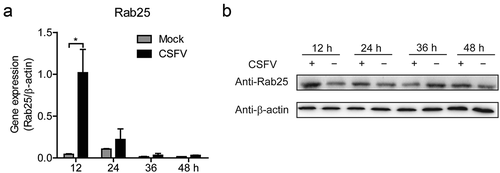
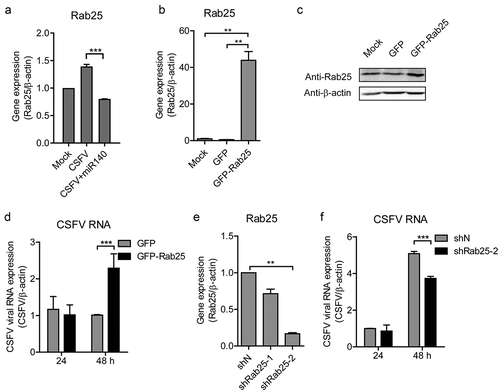
Figure 6. Rab25 promotes CSFV replication
(a) Real-time qRT-PCR analysis of Rab25 expression (normalized to β-actin expression) after transfection miR-140 mimics (150 nM) and infected with CSFV (MOI = 0.1). (b) Real-time qRT-PCR analysis of Rab25 expression (normalized to β-actin expression) after transfection of Rab25-GFP or control plasmid (GFP) for 24 h in SUVECs. (c) Immunoblot analysis of Rab25 protein levels. The number means the intensity of Rab25 protein level (top) normalized to that of β-actin. The treatment is the same as (a). (d) The SUVECs were transfected with Rab25-GFP or control plasmid and then infected with CSFV (MOI = 0.1). Real-time qRT-PCR analysis of CSFV viral RNA (normalized to β-actin expression) 24 h and 48 h post-infection. (e) The Rab25 gene was silenced by using shRNA approach. Real-time qRT-PCR analysis of Rab25 expression (normalized to β-actin expression) after transfection of shRab25 (shRab25-1, shRab25-2) or control (shN) plasmid. (f) The SUVECs were transfected with shRab25 (shRab25-1, shRab25-2) or control (shN) plasmid and then infected with CSFV (MOI = 0.1). Real-time qRT-PCR analysis of CSFV viral RNA (normalized to β-actin expression) 24 h and 48 h post-infection. Results in (A, C, D, and E) are expressed as mean ± SD of three independent experiments. p values were calculated using Student’s t-test. Western blots were analyzed and quantified using the Image J software. **P < 0.01, ***P < 0.001. NS, not significant.