Figures & data
Figure 1. Schematic diagram of yeast, pseudohypha, and hypha (black arrow indicates septin ring)
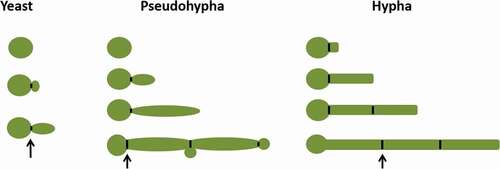
Figure 2. Cek MAPK pathway. The Cek1 mitogen-activated protein kinase pathway (MAPK pathway, dark blue) is induced by the embedded matrix environment (light blue), cell wall damage (dark blue), and low nitrogen (gray) and eventually leads to mycelium formation via the phosphorylation of transcription factor Cph1, Ace2
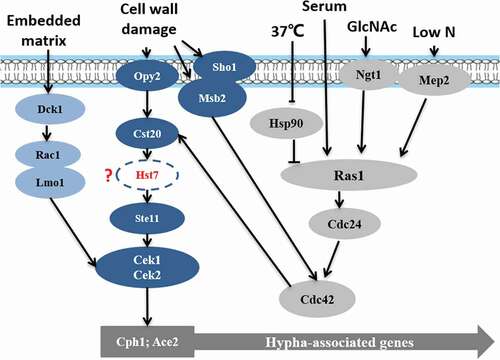
Figure 3. Regulatory models of cAMP-PKA signaling pathway in C. albicans.
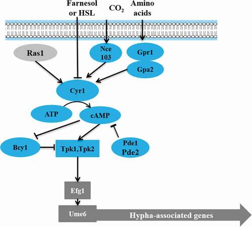
Figure 4. The Rim101‑pH sensing pathway
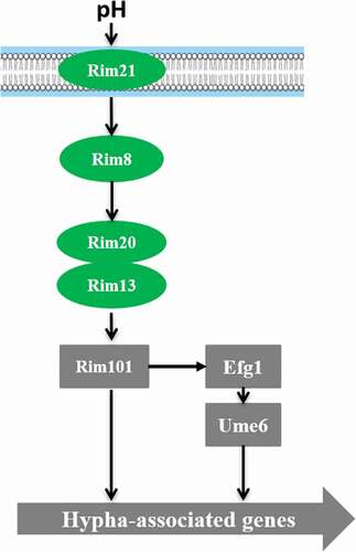
Figure 5. Hog MAPK pathway
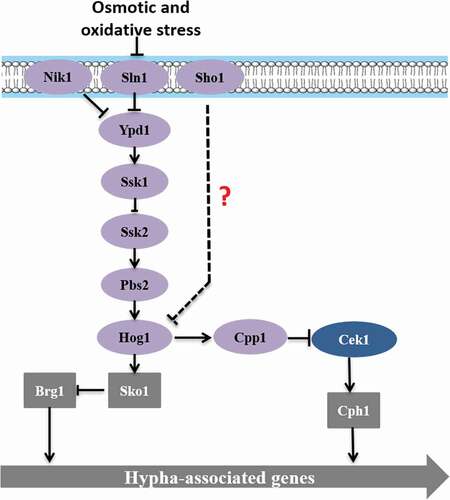
Figure 6. Tup1-mediated negative regulatory pathway
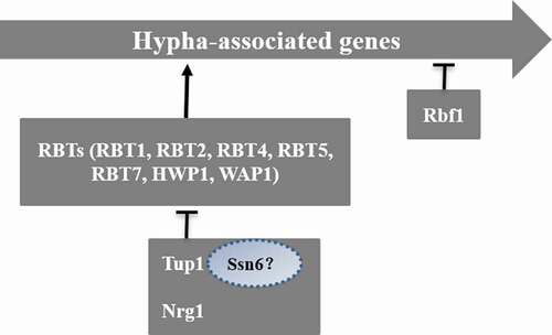
Figure 7. Key factors of mycelial elongation. Transcription factors such as Efg1 and Cph1 are involved in this regulation process. Eed1, Hgc1, Ume6 play key roles in mycelial elongation. Ume6 and Eed1 also negatively regulate Tup1 and Nrg1
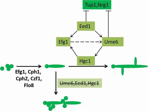
Figure 8. Transport secretory vesicles to mycelium tip. The secretory vesicles are transported to the top by actin. All vesicles will carry Sec4. After forming Spitzenkörper, the vesicles are polarized by the polar bodies and transported to the cell surface by a motorized protein strip
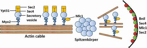
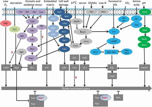
Figure 9. The pathway of hyphal formation. Different colored circles indicate different signaling pathways. The gray rectangles represent transcription factors. “?” represents that the mechanism is still uncleared here