
Figures & data
Figure 1. Phylogenetic tree illustrating the relatedness of the species included in this study. Values at nodes are SH-like local support. Scale bar indicates one substitution per 10 nucleotide positions.
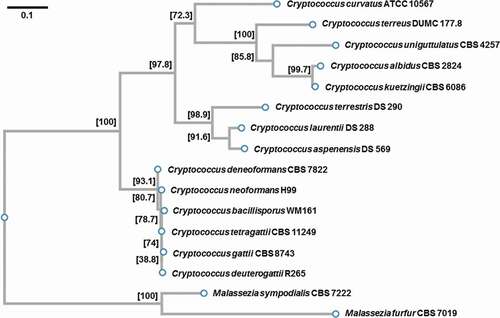
Table 1. Total number of cells and percentage of Titan cells after 48 h incubation under titanization conditions by Dambuza et al., at 30°C. The initial inoculum was 103 cells.
Figure 2. Titan cell formation among the C. neoformans and C. gattii species complex.
(a) Features of a Titan cell as observed with the DIC microscopy. C. neoformans var. grubii (strain H99) was grown under titanization conditions according to Dambuza et al. [Citation33] at 30°C for 5 days. (b) The effect of temperature (24, 30, and 37°C) on cell diameter of two C. neoformans strains (H99 and MD31) and C. deuterogattii (R265). Cells were incubated in YPD for 24 hours prior to imaging. (c) The effect of 37°C as compared to 30°C on the median cell diameter under titanization conditions [Citation33] for C. neoformans (H99, MD31) and C. deuterogattii (R265). (d) Representative images illustrating cells incubated at titanization conditions [Citation33] at 30°C, whose diameters are plotted in C. (e) The effect of incubation under titanization conditions [Citation33] at 30°C for 5 days (marked as #2) on cell body diameter of C. neoformans (H99) and representative strains of the C. gattii species complex. Cells were grown in YPD at 30°C for 8 hours as control (marked as #1). (F) Images of cells whose diameters are plotted in E (#1 – YPD control, #2 – titanization conditions at 30°C for 5 days). The number of cells counted for each condition were 100 (in b), 110 (;in c), and 167 (in e). The numbers above each plot in C and E represent the percentage of cells with the diameter above 10 μm. Statistical significance of the difference in the mean cell diameter is indicated as follows: ns (not significant), **** (p < 0.0001) Bars in (a, d), and (f) represent 10 µm.
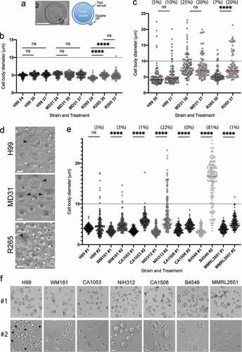
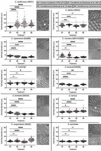
Figure 3. Members of the Cryptococcus species complex are unique in their ability to form Titan cells.
C. neoformans (MD31), and representatives of other species, as indicated, were grown in YPD control medium at 24°C (indicated as #1), subject to the in vitro titanization protocol established by Dambuza et al. [Citation33] with a modified temperature to 30°C and incubated for 48 h (indicated as #2) or 5 days (indicated as #3) or subject to conditions developed by Hommel et al. [Citation34] (indicated as #4). Cell diameters of 100 cells from each condition were measured and plotted. For each strain, representative images are shown of the cells grown at 24°C in rich YPD medium (#1) or after 48-h incubation in titanization conditions by Dambuza et el. at 30°C (#2). Statistical significance of the difference in the mean cell diameter is indicated as follows: ns (not significant), * (0.05 > p < 0.01), ** (0.01 > p < 0.001), *** (0.001 > p < 0.0001), **** (p < 0.0001). Bars represent 10 µm.
Supplemental material