Figures & data
Table 1. Oligonucleotide sequences.
Figure 1. INP0341 impedes cytotoxic effects of P. aeruginosa on HCEC. Chemical structure of INP0341 (a). HCEC were exposed to different concentrations of INP0341 for 6 h to test its effect on cell viability using the lactate dehydrogenase (LDH) assay. Cells lysed with detergent were used as a positive control and cytotoxicity was measured as a percentage of total LDH (b). HCEC were infected with PAO1 in the presence or absence of INP0341 (100 μM), or PAO1ΔpscC for 6 h and cell morphology was imaged under a bright field microscope (c) and cell viability was determined by LDH assay (d). Cells were stained with phalloidin- Alexa Fluor 488 and imaged under a fluorescent microscope using a 100x objective and oil-immersion (e). The experiments were repeated at least three times.
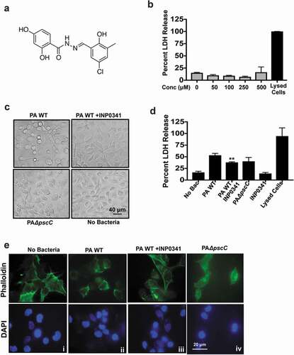
Figure 2. Increased expression of AMPs and generation of ROS in HCEC exposed to P. aeruginosa in presence of INP0341. HCEC were infected with PAO1 (a) or clinical isolates (b and c) in the presence or absence of INP0341 for 4 h, and AMP gene expression was determined by quantitative PCR using the 2−ΔΔCt method. GAPDH was used as a housekeeping gene. For detection of ROS, HCEC were exposed to PAO1 in the presence or absence of INP0341 (100 μM) or PAO1ΔpscC for 2 h and incubated with H2CFDA for 15 min and then observed and imaged using a fluorescent microscope (d). The generation of ROS was also quantitated using a fluorescent plate reader (e). The experiments were done in duplicate and repeated three times. (*indicates p < 0.05).
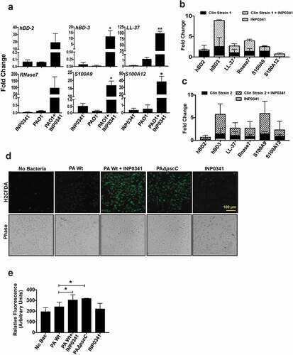
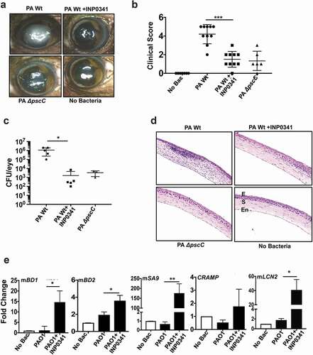
Figure 3. INP0341 attenuates P. aeruginosa infection and facilitates bacterial clearance in a murine model of keratitis. C57BL/6 mice were infected with P. aeruginosa and topically treated with INP0341 at 0, and 6 h post infection. Mice were euthanized 24 h post infection and representative images of corneal opacification (a) and their clinical score (b) were recorded. Cfu was measured from whole eye homogenates 24 h post infection (n = 5 mice). Data points represent individual infected corneas (c). Corneal sections were stained with hematoxylin and eosin to visualize cellular infiltration. E, epithelium; S, stroma; En, endothelium (d). Corneas (n = 3) were excised 24 h post infection and homogenized for RNA isolation and expression of AMPs was determined by quantitative PCR using 2−ΔΔCtmethod. GAPDH was used as a housekeeping gene. (*indicates p < 0.05; ** indicates p < 0.005).