Figures & data
Figure 1. Schematic representation of HIF-regulated pathways
HIF-1 is involved in embryonic development, apoptosis, glucose metabolism, angiogenesis and cancer metastasis, among others. HIF-2 is involved in the growth and metastasis of neuroblastomas, in the expression genes related to angiogenesis and vascular function, as well as erythropoiesis. The role of HIF-3 is less clear, but it has been reported to be involved in the regulation of erythropoietin expression and in the inhibition of activity of HIF-1 and HIF-2. Green arrows indicate upregulation of the elements indicated in the corresponding boxes.
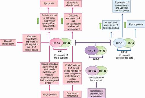
Figure 2. Schematic representation of the effects of double-stranded DNA viruses on HIF-1α and (possible) mechanisms of action
From left to right: John Cunningham virus (JCV) infection of glial cells increases the levels of HIF-1α in the nucleus; Human papilloma virus (HPV) viral oncoproteins stabilize HIF-1α; HIF-1α is stabilized in herpes stromal keratitis (HSK) by Herpes simplex virus type 1 (HSV-1) infection; Kaposi´s Sarcoma-associated herpesvirus (KSHV) upregulates HIF-1α transcription levels and stabilizes HIF-1α via its interaction with vIRF3; Human cytomegalovirus (HCMV) increases the expression and stabilization of HIF-1α; Epstein-Barr virus (EBV) induces the synthesis of HIF-1α protein and increases its mRNA levels via LMP1.
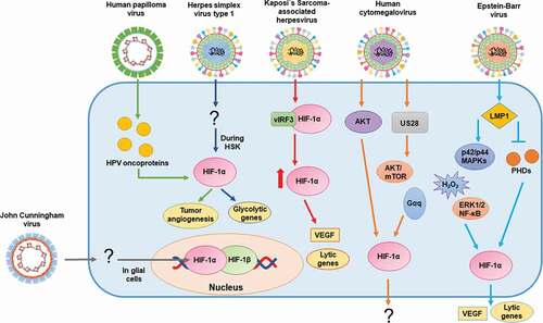
Figure 3. Schematic representation of the effects of positive single-stranded RNA viruses on HIF-1α and (possible) mechanisms of action
From left to right: Severe acute respiratory syndrome coronavirus 2 (SARS-CoV-2) causes hypoxemia. The accumulation of HIF-1α increase the levels of angiotensin II and decreases ACE2 expression; Hepatitis E virus (HEV) increases AKT/protein B and stabilizes HIF-1α; HIF-1α regulates genome replication of Dengue virus (DENV); Hepatitis C virus (HCV) stabilizes and promotes the overexpression of HIF-1α via NF-κB, STAT-3, PI3-K/AKT, p22/p44 MAPKs and the HCV core protein.
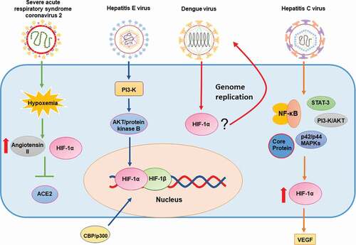
Figure 4. Schematic representation of the effects of negative single-stranded RNA and double-stranded RNA viruses on HIF-1α, and (possible) mechanisms of action
From left to right: Influenza A virus (IAV) stabilizes HIF-1α and promotes its translocation to the nucleus by inhibiting proteasome degradation and decreasing the expression of FIH-1; Human respiratory syncytial virus (hRSV) increases the expression and stabilization of HIF-1α and NO released from infected cells affects these processes; Newcastle disease virus (NDV) inhibits HIF-1α accumulation in a post-translational manner; HIF-1α increases cell resistance to Vesicular stomatitis virus (VSV) infection. Mammalian orthoreovirus (MRV) downregulates HIF-1α likely through endo-lysosomal escape of viral genome into the cytoplasm.
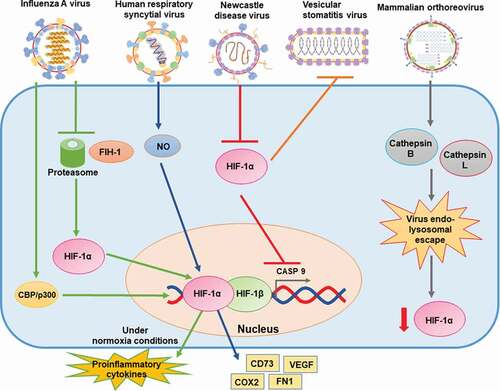
Figure 5. Schematic representation of the effects of retrotranscribing viruses on HIF-1α and (possible) mechanisms of action
From left to right: Human immunodeficiency virus (HIV) promotes HIF-1α expression and stabilization via Vpr protein and mitochondrial ROS; Hepatitis B virus (HBV) increases the transcriptional activity and protein levels of HIF-1α. The HBV HBx protein may inhibit pVHL binding to HIF-1α.
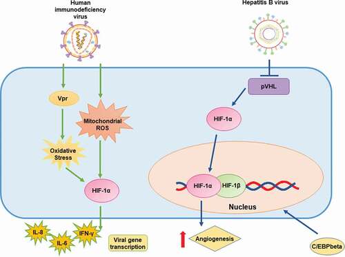
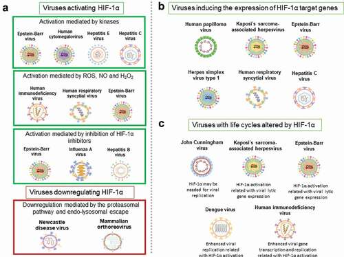
Figure 6. Common features between different viruses and HIF-1α. A
Viruses activating or downregulating HIF-1α. Viruses activating HIF-1α via kinases: EBV, HCMV, HEV and HCV. Viruses activating HIF-1α via ROS, NO and H2O2: HIV, hRSV and EBV. Viruses activating HIF-1α via the inhibition of HIF-1α inhibitors: EBV, IAV and HBV. Viruses downregulating HIF-1α via the proteasomal pathway and by endo-lysosomal escape: NDV and MRV. B Viruses inducing the expression of HIF-1α target genes: HPV, KSHV, EBV, HSV-1, hRSV and HCV. C Viruses with life cycles altered by HIF-1α: JCV, KSHV, EBV, DENV and HIV.