Figures & data
Figure 1. Mechanisms of JEV neuroinvasion. The numbers in squares indicate the mechanisms of virus entry into the brain: 1, passive transport of virus particles across the endothelial cells; 2, diapedesis of infected leukocytes; 3, virus transport via the peripheral nervous system; and 4, virus transport through the BBB disrupted by inflammatory mediators released from cells of blood and brain sides of the BBB. The inflammatory mediators written in gray mediate the crosstalk between microglia, astrocytes, and neurons that may contribute to the BBB damage. The symbol “?” denotes the missing information in the literature. Created with the web-based BioRender tool (BioRender.com)
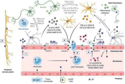
Figure 2. Proteins-mediated active inflammatory responses during JEV infection. The symbol “?” denotes the unidentified upstream regulator. Created with the web-based BioRender tool (BioRender.com)
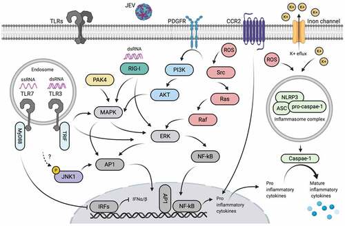
Figure 3. Non-coding RNA-mediated active inflammatory responses during JEV infection. Created with the web-based BioRender tool (BioRender.com)
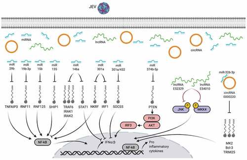
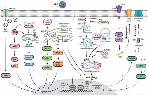
Figure 4. Mechanisms of neuronal cell damage during JEV infection. The symbol “?” denotes the cellular processes that have been confirmed in JEV-infected peritoneal macrophages, but not in neurons. Created with the web-based BioRender tool (BioRender.com)