
Figures & data
Figure 1. Identification of the region of Tip critical in PilB inhibition
(a) Schematic representation of the truncated Tip derivatives and their activities. The deletions at the N-terminus (NΔ60, NΔ80, and NΔ100) and at the C-terminus (CΔ20 and CΔ40) from Tip were constructed to determine the regions critical for the Tip activity. They are either deficient (-) or proficient (+) in the Tip activity (PilB interaction and twitching inhibition) as shown in b and c. The critical regions for the Tip activity are designated in blue colors with the amino acid numbers. (b) Bacterial two-hybrid assay with the truncated Tip derivatives was performed to determine the PilB-binding region. The full-length (FL) Tip and the truncated derivatives in A were cloned into pKT25 and then introduced to E. coli DHP1 containing pUT18C-PilB. The cells with Tip-PilB interaction were examined by β-galactosidase activity. (c) Twitching motility was assessed using the PA14 cells harboring the full-length (FL) Tip and the N-terminally truncated Tip derivatives in a, which were introduced at the attTn7 site as described elsewhere [Citation21]. Twitching motility was examined in the absence (-) or presence of 1 mM IPTG.
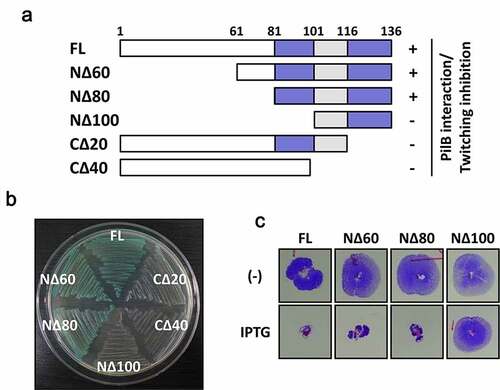
Figure 2. Design and synthesis of a series of Tip-derived peptides
(a) Partial sequence alignment between Tip and its homologs of the related phages. A series of 6 peptides (P1 to P6) were designed with the spanned regions designated as colored horizontal lines. Each peptide consisted of 15–16 aa long with a 5-6-aa overlap between the neighboring peptides in a row. The critical regions for the Tip activity are designated in blue colors as in ). (b) Chemical synthesis of the Tip-derived peptides in (a). The sequences of the 6 peptides are shown with C-terminal amidation. The underlined residues (red and green) of P1 denote those subjected to the point mutation analysis (see texts and ). Asterisk (*) indicates the discarded peptide due to its poor aqueous solubility (P5).
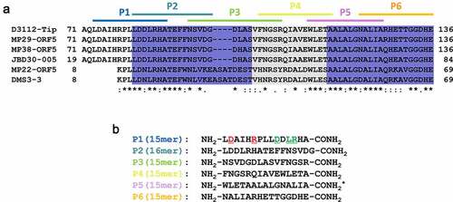
Figure 3. Bioactivity of the Tip-derived peptides upon exogenous administration
(a) Interference with TFP-specific phage infection. Ten and 100 plaque forming units (pfu) of PP7 and MPK7 phages [Citation23,Citation32] were mixed with the synthetic Tip-derived peptides (5 mM) in , except for P5 with poor solubility. The phage-peptide mixtures were spotted onto the lawn of PAO1. The TFP inhibition activities were assessed by the peptide-mediated reduction of pfu. (b) Interference with PilB-Tip interaction. E. coli stain, DHP1 containing pUT18C and pKT25 (vector), pUT18C-Zip and pKT25-Zip (Zip/Zip), and pUT18C–PilB and pKT25-Tip (PilB/Tip) were incubated with either nothing (-) or one of the synthetic peptides (P1, P2, and P6) for 10 min and spotted on a MacConkey agar supplemented with 1% maltose. (c) Twitching inhibition. Five microliters of either TDW (-) or one of the synthetic peptides (5 mM) were directly treated to the PA14 colonies on LB agar for 10 min, and then the colonies were used for twitching motility assay. The twitching zones were stained with 0.1% crystal violet. (d) PilB delocalization. The PA14 cells containing PilB fused with GFP [Citation21] was treated with either TDW (-) or one of the synthetic peptides (1 mM) for 10 min. PilB delocalization was monitored by fluorescence microscope as described in Methods. The polar (i.e. both unipolar and bipolar) localization of PilB in the cells was indicated as a red arrow. (e) Quantitation of PilB delocalization. Each fraction of PilB localization (polar, empty; diffuse, filled) in the independent fields was measured and represented with error bars represent the standard deviations. Statistical significance between the groups is indicated, based on a p value of less than 0.001 (***) by Student’s t test.
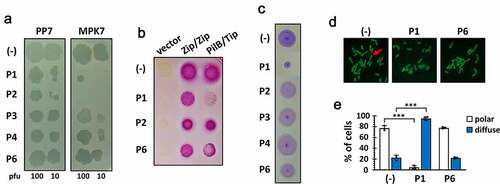
Figure 4. Structure and membrane permeability of P1
(a) Helical wheel projection of P1. The hydrophobic and hydrophilic residues are separated by a dotted line. The arrows indicate the helical moment. Amino acid color codes: hydrophobic, yellow; cationic, dark blue; anionic, red; alanine and glycine, gray; proline, green; histidine, light blue. (b) Fluorescence quenching of the FITC-P1 conjugates by liposome. The liposome composed of phosphatidylethanolamine, phosphatidylglycerol and cardiolipin was prepared to mimic the P. aeruginosa membrane. FITC-P1 (64 μM) was incubated with noting (-; empty) or the liposome (+; filled). Triton-X100 was added to the mixture after 60 min (arrow). Fluorescence from free (i.e. untrapped) FITC-P1 was observed at the designated time points. (c) FACS analysis of the PA14 cells after FITC-P1 treatment. PA14 cells were incubated with noting (red) or FITC-P1 (64 μM) (green) for 30 min, and then washed with PBS. Fluorescence intensity of the cells was analyzed by flow cytometry, as described in Methods.
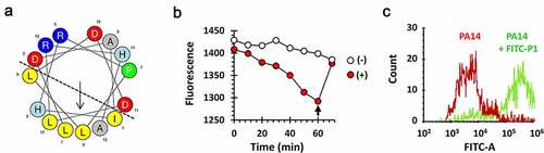
Figure 5. Amino acid residues in P1 critical for the Tip function
Twitching motility (a) and PilB localization (b) were assessed using the PA14 cells harboring the wild type (WT) Tip and its point mutants (D74K, R78G, D82K, L84P, and R85D) for the amino acids within the P1 region. Twitching motility was examined in the absence (-) or presence of 1 mM IPTG as in ) and PilB localization was monitored in the presence of 1 mM IPTG by fluorescence microscope as in ). The polar (i.e. both unipolar and bipolar) localization of PilB in the cells was indicated as a red arrow. (c) Quantitation of PilB localization. The percentage of polar localization of PilB was determined in the absence (empty) or presence (filled) of 1 mM IPTG, with the error bars representing the standard deviations. Statistical significance between the groups is indicated, based on a p value of less than 0.001 (***) by Student’s t test.
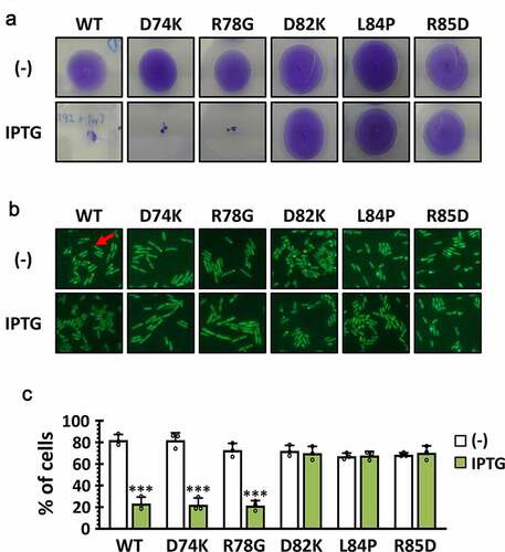
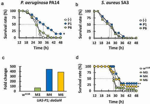
Figure 6. Antibacterial efficacy of P1 in vivo
(a) and (b). Mortality of PA14 and SA3-infected flies fed with P1. The wild-type Oregon R flies were infected with PA14 or SA3 cells that had been co-treated with nothing (empty) or 10 mM of the synthetic peptide P1 or P6 (filled). The survival rates were determined over time for the infected flies up to 48 h post-infection. (c) Ectopic P1 expression in transgenic flies. The constitutive transcriptions of P1 in UAS-P1/daGAL4 fly lines (M3, M4 and M6), which are isogenic mutants of w1118, were measured by RT-RTi PCR using total RNA extract, as describe in Methods. The relative transcriptional fold changes of P1 were determined in comparison to their parental strain w1118 and normalized using the reference gene rp49. (d) Mortality of ectopic P1-expressing transgenic flies upon PA14 infection. The w1118 (empty) and its isogenic UAS-P1/daGAL4 fly lines (M3, M4 and M6) (filled), which constitutive overexpresses P1, were systemically infected by PA14 pricking. Those infected flies were transferred to the new corn meal media and the survival rates were measured up to 48 h post-infection. The dotted lines represent the time required to reach 50% mortality. The statistical significance based on a log-rank test is indicated (***, p< 0.001).
Supplemental material