
Figures & data
Figure 1. Domain structures of S. cerevisiae core septins. All septins contain a GTP-binding domain, the septin unique element (SUE) and a phosphoinositide-binding polybasic region (PBR). The C-terminus (coiled-coil) regions vary
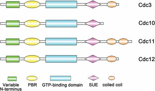
Table 1. Septins and their functions in fungi
Figure 2. Phylogeny and evolutionary history of septins. Maximum likelihood phylogenetic tree of 65 septin proteins from 11 fully sequenced species that are reported in eukaryotic lineages. The categorization of different septins by multiple copies of coiled coils found in a single protein: the Cdc11 group with two coiled-coil domains, the Cdc3 and Cdc12 groups with one coiled-coil domain and the Cdc10 group with no coiled-coil domain
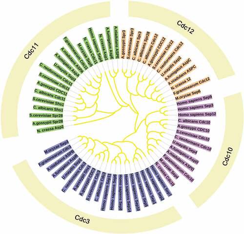
Figure 3. Structures of the septins. (a) The predicted structures of different septins in yeast cells. (b) Schematic diagram of the yeast septin rod, filament, ring, and gauze. The yeast hetero-octameric septin complex is a linear rod with the subunits arraying in the order and with the interfaces indicated. Each septin forms associations with its neighbors through either a G interface or an N-C interface. The rod septins can form high-order structures, like filaments, rings, gauzes
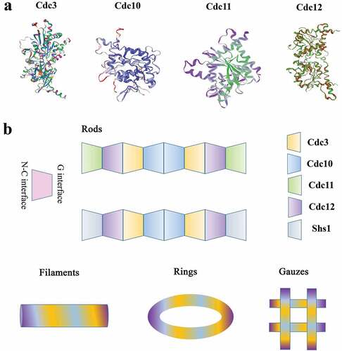
Figure 4. Model showing that septins control conidia autophagy degradation and participate in the development of the penetration peg in Magnaporthe oryzae by analogy with Saccharomyces cerevisiae. Autophagy begins after conidia are induced on the hydrophobic membrane for 12 hours. Under the induction of starvation, septin colocalized with ATG8 (A8) and ATG9 (A9) at the PAS site. Septins are involved in the biosynthesis of autophagosomes. The atypical ring formed by septins is approximately the same diameter as the autophagosome ring in M. oryzae. During the formation of the penetration peg 24 hours after induction, septins aggregated in the appressorium pores, recruited Gin4, activated Gin4 recruited Hsl7, Hsl7 recruited Swe1, and Swe1 was subsequently highly phosphorylated and degraded, and the appressorium entered the mitotic progress. (D represents degradation; C represents cell cycle)
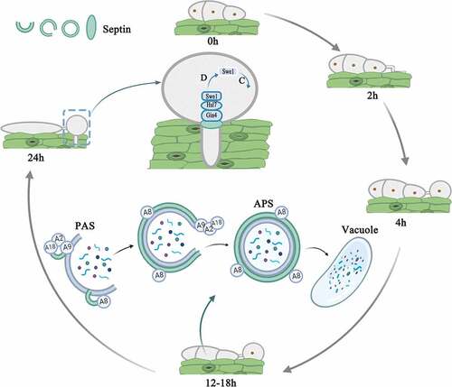
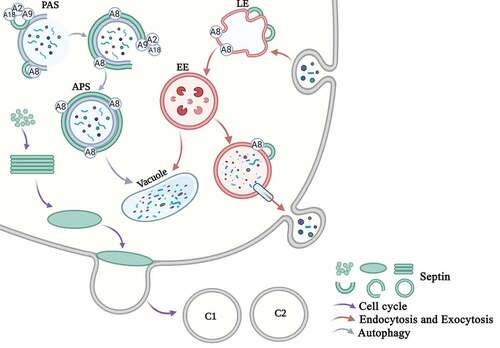
Figure 5. Overview of the main functions of septins in eukaryotic cells in S. cerevisiae. Cell cycle. Septins are mainly involved in the cell cycle progress. Free septins are recruited and aggregated into filaments, which then form a ring in the G1 phase and a collar in the S phase. The M-phase mother cell divides to form two daughter cells, and the septin ring also divides into the two daughter cells (C1 and C2). Endocytosis and Exocytosis. Septins are involved in the processes of endocytosis and exocytosis. The early endosomes swallow the extracellular proteins that enter the cell, and Sep3 and Sep6 bind to the early endosomes (EE). The late endosomes (LE) enter the vacuole and are degraded on the one hand, and they are secreted out of the cell through the vesicle on the other hand. The SNARE protein and the septin protein assists vesicle exocytosis. Autophagy. Septins are involved in autophagy. Septins are recruited to the PAS site. Sep4 interacts with Atg8 (A8), Atg9 (A9), and Atg18 (A18) and surrounds the autophagosomes (APS). Septins participate in the transport of Atg9 and the biosynthesis of the autophagosome
Data availability statement
Data sharing not applicable-no new data generated. Data sharing is not applicable to this article as no new data were created or analyzed in this study.