Figures & data
Table 1. Phenotypic and genotypic characteristics of K. pneumoniae strains used in this study
Figure 1. The hypermucoid phenotype of K. pneumoniae is associated with inhibition of cell adherence and internalization. Mucoviscosity (a) and uronic acid production (b) in hypervirulent Klebsiella strains HvKP1, HvKP2, HvKP3, HvKP4, and classical Klebsiella strains cKP1, cKP2, cKP3, cKP4. Invasion assay of selected clinical K. pneumoniae strains using Macrophage RAW 264.7 cells (c) and intestinal epithelial Caco-2 cells (d). Adhesion assay of selected strains using lung epithelial A549 cells (e) and Caco-2 cells (f). Data were analyzed by one-way ANOVA test. Each data point was repeated three times (n = 3). Data are presented as the mean ± s.e.m. * P < 0.05; ** P < 0.01; ***P < 0.001; **** P < 0.0001. Abbreviation: NS, not significant
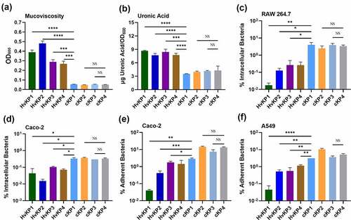
Figure 2. Cell adherence and internalization assay of K. pneumoniae strain NP-HvKP variants and HvKP5. Mucoviscosity (a), and uronic acid production (b) of Klebsiella strain NP-HvKP, NP-HvKP/A, NP-HvKP/A2, NP-HvKP-PC, NP-HvKP-TC and HvKP5. Assay of the ability of selected strains to adhere to lung epithelial A549 cells (c) and Caco-2 cells (d). Invasion assay of selected clinical K. pneumoniae strains using Macrophage RAW 264.7 cells (e) and intestinal epithelial Caco-2 cells (f). Data were analyzed by one-way ANOVA test. Each data point was repeated three times (n = 3). Data are presented as the mean ± s.e.m. * P < 0.05; ** P < 0.01; **** P < 0.0001. Abbreviation: NS, not significant
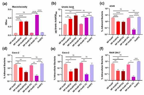
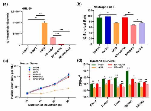
Figure 3. HvKP strains escape neutrophil-mediated phagocytosis and killing. (a) Neutrophil-like cell dHL-60 phagocytosis assay of HvKP1, HvKP2, cKP2, NP-HvKP, NP-HvKP/A, and NP-HvKP/A2. (b) Neutrophil killing assays of Klebsiella pneumoniae strains. (c) The resistance of K. pneumoniae strains to killing by pooled human serum. (d) Bacteria loads in blood, liver, lungs, kidney, and spleen were measured by plating at 24 hr post-infection. Bacterial load detectable in blood and various organs: HvKP1, cKP2, NP-HvKP/A and NP-HvKP. Data were analyzed by one-way ANOVA test. Each data point was repeated three times (n = 3). Data are presented as the mean ± s.e.m. * P < 0.05; ** P < 0.01; *** P < 0.001