
Figures & data
Figure 1. Phylogenetic relationship of pegivirus. The phylogenetic tree was constructed based on RdRp gene sequences of selected Flaviviridae members using the maximum-likelihood (ML) method (MEGA 7.0.26). Four genera are classified in the Flaviviridae family. The main hosts of these viruses are also shown in the figure. The red branches highlight the members of the genus Pegivirus.
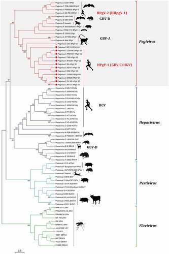
Figure 2. Genome organization of HPgV-1 and HCV. The genome encodes a single pre-polyprotein that is cleaved into mature viral proteins after co-translation and post-translation. Compared to HCV, HPgV-1 genome encodes two additional predicted proteins (protein X at upstream of E1, and protein p* between E2 and NS2), but does not encode a core protein that is an RNA-binding protein and forms the virion nucleocapsid.
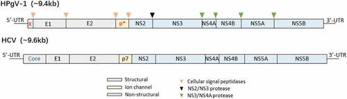
Table 1. HPgV-1 proteins and their functions
Figure 3. Global prevalence and distribution of HPgV-1. BD: blood donors; HR: high-risk population mainly including IDUs, CSWs, and MSM.
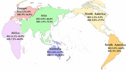
Table 2. HPgV-1 prevalence in persons co-infected with HIV-1 or HCV or EBOV and clinical outcomes
Data Availability Statement
Data sharing is not applicable to this article as no new data were created or analyzed in this study.