
Figures & data
Figure 1. HK-C. albicans primed BMDMs show increased antibacterial functions. a) Chronogram of the BMDM experiment. C57BL/6 WT mice were injected IP with an inducer of trained immunity in 100 μL before bone marrow cells were collected. Control mice were injected IP with the same volume PBS. Bone marrow cells were differentiated into bone marrow-derived macrophages (BMDMs) for six days, plated, and stimulated with Clostridium perfringens at a MOI of 5 or PBS vehicle. b) the killing ability of BMDM primed with PBS or HKCA (HK-C. albicans). 3 h post C. perfringens (MOI = 5) infection, viable bacteria were plated on BHI agar to determine CFU counts.

Figure 2. Trained BMDMs increased release of extracellular traps. a) Extracellular DNA release from BMDM cultures was quantified. The release of extracellular DNA from BMDM (primed with PBS, 1 × 10⁶ CFU and 5 × 10⁶ CFU heat-killed C. albicans), was detected by SYTOX Green 2 h,3 h,4 h post-stimulation with the C. perfringens (MOI = 5). Extracellular DNA from cells was incubated without C. perfringens assumed as 1. Results are shown as fold increase relative to cells without C. perfringens. b) Western blot analysis of protein expression. Non-primed and HK-C. albicans-primed BMDMs were infected with C. perfringens (MOI = 5) or not for 3 h, whole cell lysates were analyzed for CitH3, PAD4 and GAPDH by Western blotting.
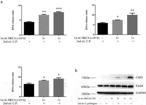
Figure 3. Mannan-Induced BMDM enhanced the memory response of ETs. a) Extracellular DNA release from BMDM cultures was quantified. BMDMs from non-primed and mannan-primed (1 mg/ml) mice were infected with C. perfringens (MOI = 5) or not. 3 h post-infection, the extracellular DNA in cell supernatants was stained with SYTOX Green and detected with a fluorescent reader. BMDMs incubated without C perfringens were used as control, assumed 1. Results are shown as fold increase relative to cells without C. perfringens. b) Western blot analysis of protein expression. Non-primed and mannan-primed BMDMs were infected with C. perfringens (MOI = 5) or not for 3 h, whole cell lysates were analyzed for CitH3, PAD4 and GAPDH by Western blotting.
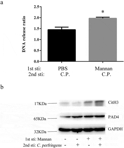
Figure 4. HK-C. albicans leads to trained memory of ETs in chicken PBMC. a) Chronogram of the chicken PBMC experiment. the chickens have been injected IP with 1 × 10⁶ CFU heat-killed C. albicans in 100 μL before the collection of PBMCs. Adherent cells were second stimulated with Clostridium perfringens or PBS vehicle at the designed time points. b) the release of extracellular DNA from HK-C. albicans-primed PBMCs was detected by SYTOX Green 3 h post-stimulation with the Clostridium perfringens (MOI = 5) or not. PBMCs incubated without C perfringens were used as control, assumed 1. Results are shown as fold increase relative to cells without C. perfringens. c) the killing ability of PBMCs. The extracellular bacteria of PBMC 3 h post C. perfringens (MOI = 5) infection was assessed by plated on a BHI agar plate. d) PBMCs nitric oxide (NO) production. Non-primed and HK-C. albicans primed PBMCs infected with C. perfringens (MOI = 10), 24 h post-infection, the cell culture supernatants were collected and nitric oxide was measured by Griess reaction. PBMCs incubated without C perfringens were used as a control (data not shown).
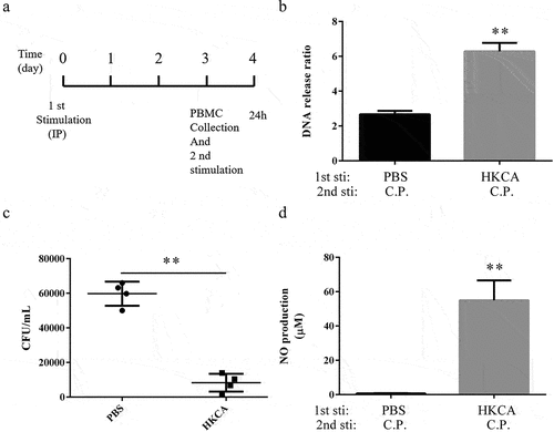
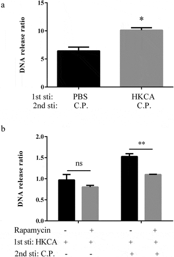
Figure 5. Rapamycin inhibits the trained memory of ETs. Intraperitoneal injection in C57BL/6 WT mice with 4 mg/kg rapamycin for 30 min prior to HK-C. albicans (1 × 10⁶ CFU) primed. Animals were sacrificed 3 days after injection and collected BMDMs. BMDMs infected with C. perfringens (MOI = 5) or PBS vehicle and fluorescence of extracellular DNA was measured 3 hours after infection. a) Extracellular DNA release was quantified. BMDMs stimulated with PBS were used as control. Fold changes in DNA release obtained in BMDM after stimulation with C. perfringens. the ratio of DNA release by C. perfringens versus PBS stimulation BMDMs is presented. b) Non-primed BMDMs infected without C. perfringens was used as control, was assumed to be 1. DNA release was quantified and presented as ratio vs control.
Supplemental Material
Download Zip (142.7 KB)Data availability statement
The authors confirm that the data supporting the findings of this study are available within the article and its supplementary materials.