
Figures & data
Figure 1. Construction and characterization of plasmids. (a) the strategy for constructing the infectious plasmid clone pJN containing the entire genome of the NGPV strain SDJN19. (b) Illustration of pJN harboring the SDJN19 genome. (c) Characterization of pJN by restriction endonuclease digestion. 1,2: Digestion with SphI produced 3.3 kb, 2.5 kb and 2.2 kb DNA fragments; 3,4: Double digestion with XhoI and BamHI produced a 3.0 kb vector and 5.0 kb genomic molecule. (d) the diagram depicting construction of the chimeric viruses. the original Rep1 or VP1 gene in plasmid pJN was replaced by the counterpart from the classical GPV strain LH through overlap PCR and usage of suitable restriction endonuclease sites.
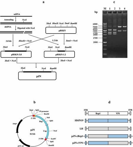
Figure 2. Influence of rJNm infection on body weight, beak development and survival of Cherry Valley Pekin ducks. (a) Measurement of body weight at 7, 14, 21, 28 days post-infection of 2-day-old ducks. (b) Measurement of beak length. (c) Measurement of beak width. ****, p < .0001, significant difference between the infection group and control group. (d) Death numbers of ducks observed at different days post-infection.
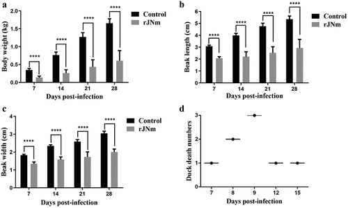
Figure 3. The gross clinical signs of Cherry Valley Pekin ducks at different days post-infection of rJNm. These signs included the inability to stand and move, lameness, one or both legs stretching back or outwards, protruding tongues, delayed molt (A-E), red and swollen metatarsus (F).
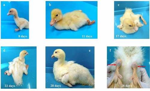
Figure 4. Gross lesions observed in the dead ducks after rJNm infection. Congestion was widely found in the internal organs, including heart, spleen, liver, lung, kidney, pancreatic gland (A-E, G-H). The gallbladder was full of dark green bile (E). The glandular stomach surface (F) and the esophageal mucosa (I) had excessive thick mucus attached. The duodenum was dilated and relaxed (J), and the mucosa became thinner, pale red, and had yellow inflammatory secretions attached (K).
Figure 5. Viral loads in the internal organs of the dead ducks were determined by qPCR, which were compared with that of the deceased goslings after infection with the classical GPV strain LH. ***, significantly different, P<0.001.
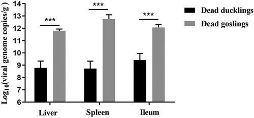
Figure 6. X-ray and micro CT analysis of duck bone tissues. (a) X-ray imaging of duck legs of the living ducks at 28 dpi. left, the rJNm-infected duck; right, the control duck. (b) X-ray imaging of duck wing. above, the rJNm-infected duck; below, the control duck. (c) Coronal section images of tibias of the living ducks at 21 dpi were created by micro CT. left, the control duck; right, the rJNm-infected duck. (d) Comparison of BV/TV, SMI and BMD of the tibias between the infected and control ducks at 21 dpi. *, significantly different, P < 0.05.
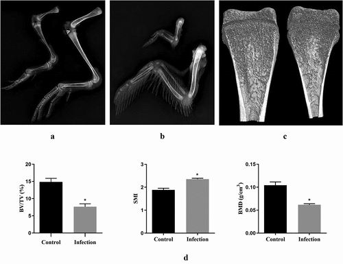
Figure 7. Performance of Cherry Valley Pekin ducks infected with rJNm via the horizontal transmission route. A-C: Comparison of duck body weight, beak length, beak width among the control group, the horizontal contact group and the rJNm infection group. *, p < 0.05; **, p < 0.01; ***, p < 0.001; ****, p < 0.0001; ns, no significant difference (p > 0.05). D: Tongue protrusion was clearly observed in the horizontal contact group ducks. E: Viral loads comparison of the internal organs between the dead ducks in the rJN infection group and the living ducks in the horizontal contact group at 31 days post-infection (dpi). F: 800-bp DNA fragments covering the genetic marker site were amplified from the spleens, ileums, but not livers of the living ducks in the horizontal contact group at 31 dpi. 1, 4: liver; 2, 5: spleen; 3, 6: ileum. G: Sequence analysis of the amplified 800-bp fragments revealed common nucleotide mutations (A→G) present in the dead ducks from the rJNm infection group and the living ducks from the horizontal contact group. H: Serum antibody titres of the living ducks in the horizontal contact group at 30 dpi were measured by the agar gel precipitation test and compared with the infection group. ns, no significant difference, p>0.05.

Figure 8. Comparison of body weights, beak lengths and other signs between therJN-cRep1 and the rJN-cVP1 infected ducks. (A-B) Comparison of body weights and beak lengths among the chimeric virus infection groups and the control group. *,p<0.05; **, p < 0.01; ***, p < 0.001; ****, p < 0.0001; (C) Two ducks in the rJN-cRep1 infection group showed red and swollen metatarsus at 28 dpi. (D-F) Ducks in the rJN-cVP1infection group showed typical signs of SBDS at 28 dpi, including dwarfishbody, tongue protrusion, lameness, delayed molt, and red and swollen metatarsi.
Table 1. Comparison of the clinical signs of Cherry Valley Pekin ducks post-infection of chimeric viruses
Supplemental Material
Download Zip (409 KB)Data availability statement
The datasets generated and/or analyzed during the current study are not publicly available due to the project is not finished yet but are available from the corresponding author on reasonable request.