
Figures & data
Figure 1. EHEC hemolysis regulation by CsrA.
a, Hemolytic effect of EhxA on EHX blood agar plate: 1, EHEC wild type; 2, ΔcsrA; 3, ΔcsrA/pCSRA; 4, ΔcsrA/ΔhlyE; 5, ΔcsrA/ΔehxA; 6, ΔcsrA/ΔehxB; 7, ΔcsrA/ΔehxB/pEHXB; 8, ΔcsrA/ΔehxB/pACYC184. b, EhxA secretion level of EHEC strains. Western-blotting analysis for the supernatant and pellet of EHEC culture. The target protein EhxA was added a His-tag at its N-terminus. The strains were transformed into pQE-ehxA and pCSRA. Empty plasmids pQE80L and pACYC184 were used as negative controls. c, Hemolytic effect of plasmid-copy HlyE on EHX blood agar plate.
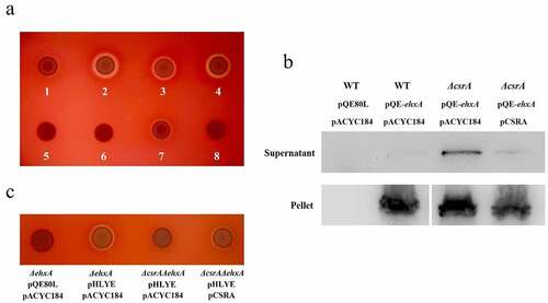
Figure 2. Overview of ehxCABD (EHEC), hlyCABD (UPEC), and hlyE (EHEC) operons.
The DNA sequences contain non-coding regions (blue), 30 bp of the 5’ and 3’ ends of gene coding regions (black), and GGA motifs (red). The secondary structure of underlined DNA sequences, predicted using CentroidFold, is displayed on the right side of each corresponding DNA sequence. a, ehxCABD; b, hlyCABD; c, hlyE.
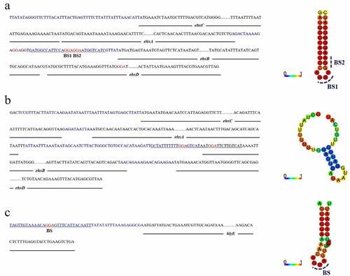
Figure 3. CsrA-Dependent regulation of ehxB-lacZ, hlyE-lacZ, and ehxA-lacZ translational fusion in vivo.
Bacterial β-galactosidase activity was detected at various time points throughout growth. Beta-galactosidase activity (green triangle: wild-type; orange triangle: ΔcsrA) and corresponding bacterial growth (blue square: wild-type; red circle: ΔcsrA) were determined for EHEC. a, ehxB-lacZ translational fusion; b, hlyE-lacZ translational fusion; c, ehxA-lacZ translational fusion. Each value represents the mean ± SD of three independent measurements. The significance of β-galactosidase activity in ΔcsrA compared with WT is also indicated: *p< 0.05, **p< 0.01, ***p< 0.001.
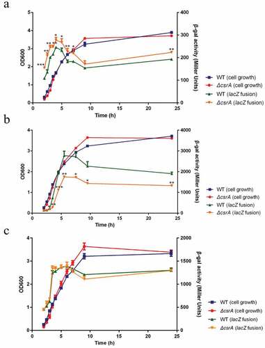
Figure 4. RNA mobility shift assays using various concentrations of purified 6hisCsrA on EHEC.
The concentration of CsrA in lanes 1 to 10 was 0, 0.9, 1.8, 3.6, 7.2, 14.4, 28.8, 43.2, 57.6, and 72 nM, respectively; the concentration of the FAM-labeled probe was 1 μM. a, interaction between 6HisCsrA and FAM-labeled positive control transcript R9-43; b, interaction between 6HisCsrA and FAM-labeled negative control transcript hns; c, interaction between 6HisCsrA and FAM-labeled transcript of ehxB (ehxB-Ori); d, interaction between 6HisCsrA and FAM-labeled transcript of hlyE (hlyE-Ori); e, interaction between 6HisCsrA and FAM-labeled transcript of ehxB with BS1 motif mutation (ehxB-MuBS1); f, interaction between 6HisCsrA and FAM-labeled transcript of ehxB with BS2 motif mutation (ehxB-MuBS2); g, interaction between 6HisCsrA and FAM-labeled transcript of ehxB with BS1 and BS2 motif double mutation (ehxB-MuBS1-2); h, interaction between 6HisCsrA and FAM-labeled transcript of hlyE with CsrA binding motif mutation (hlyE-Mu). B, bound RNA; F, free RNA.
Figure 5. Effect of site-directed mutation in potential binding sites in the expression of lacZ translational fusion in vivo.
a, Schematic diagram of translational fusion constructs for β-galactosidase assays; b, ehxB-lacZ translational fusion detected by the ONPG method. Strains were cultured in LB medium for 5 h at 37 ℃ under constant shaking at 250 rpm; c, hlyE-lacZ translational fusion detected by the ONPG method. EHEC was cultured in LB medium for 5.5 h at 37 ℃ under constant shaking at 250 rpm. Significantly different β-galactosidase activity is indicated: **p< 0.01, ***p< 0.001, ****p< 0.0001.
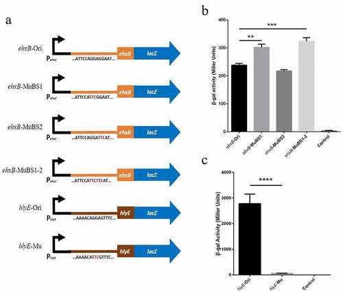
Figure 6. Stability of ehxB and hlyE mRNA as affected by CsrA in EHEC.
EHEC wild-type and ΔcsrA cells were cultured in LB medium to OD600 = 0.4, treated with 200 μg/mL rifampicin, and collected at the indicated time points after treatment. Total RNA was extracted using the RiboPureTM Bacteria kit, and the purified RNA was used for cDNA synthesis by reverse-transcription PCR. The cDNA was then used as template for further amplification. a, ehxB mRNA detection; b, hlyE mRNA detection. Amplifications were carried out for 29 cycles (ehxB) or 30 cycles (hlyE), and each product was analyzed by agarose gel electrophoresis. c, mRNA degradation ratio detection by qPCR. Dashed line: ehxB; green triangle: ehxB in wild-type; orange triangle: ehxB in ΔcsrA; solid line: hlyE; blue circle: hlyE in wild-type; red square: hlyE in ΔcsrA. 16S rRNA was used as the reference for normalizing the target products. Each value represents the mean ± SD of three independent measurements.
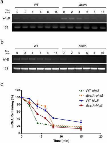
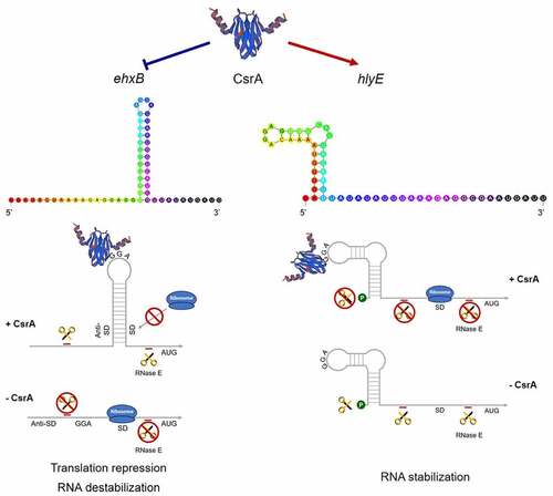
Figure 7. CsrA dual regulation of EHEC hemolysis.
Hemolysis regulation by CsrA was predicted in EHEC. The 5′ UTRs of both ehxB and hlyE can form well matched stem-loop structures with GGA motifs. CsrA directly binds to the RNA leader sequence of ehxB to repress its expression in two different ways: CsrA either binds to the SD sequence of ehxB to block ribosome access or to the ehxB transcript to promote its mRNA decay. It can also interrupt EhxA secretion and downregulate ehxA-derived hemolysis. Binding of CsrA to the hlyE transcript may stabilize its mRNA and promote hlyE-derived hemolysis.
Supplemental material
Supplemental Material
Download Zip (66.9 KB)Data availability statement
The authors confirm that the data supporting the findings of this study are available within the article and its supplementary materials.