
Figures & data
Figure 1. Geographical distribution of 273 Shewanella infectious cases. The geographical distribution of cases are Denmark (n = 71), Spain (n = 39), Africa (n = 30), China (n = 19), U.S.A (n = 17), Martinique (n = 16), India (n = 12), France (n = 9), Japan (n = 7), Korea (n = 5), Turkey (n = 6), Australia (n = 5), Caucasian (n = 3), Croatia (n = 2), Malaysia (n = 2), Thailand (n = 2), Belgium (n = 2), Italy (n = 2), Panama (n = 1), Mexico (n = 1), Moroccan (n = 1), Belize (n = 1), Wakefield (n = 1), Virginia (n = 1), Bahamas (n = 1), Romania (n = 1), Madagascar (n = 1), Germany (n = 1), Caribbean (n = 1), Cyprus (n = 1), UK (n = 1), Brunei Darussalam (n = 1), Puerto Rico (n = 1), Russia (n = 1), New Zealand (n = 1), Côte d’Ivoire (n = 1), and Israel (n = 1). Information on the geographical location of 5 cases is not available.
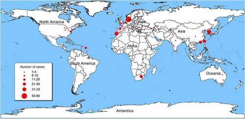
Table 1. Clinical characteristics associated with Shewanella infection.
Figure 2. Clinical infections and diseases caused by Shewanella species.
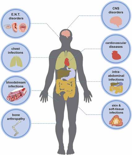
Table 2. Summary report of the underlying diseases of Shewanella infection.
Figure 3. Concatenated split network tree based on six genes loci. The gyrA, gyrB, infB, recN, rpoA, and topA gene sequences from 66 Shewanella species were concatenated and reconstructed using SplitsTree program (version: 4.14.4), based on Jukes-Cantor model.
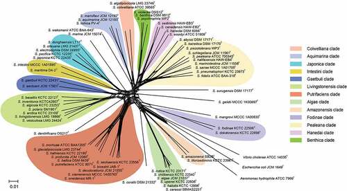
Table 3. The treatment regimen and drug resistance of strains summerized in the case reports.
Figure 4. Colony morphology of Shewanella strains on different plates. Figure 4a, 4b, 4c and d showed the colonies on marine agar 2216, nutrient agar, TCBS and MacConkey agar, respectively, after incubating at 37 °C for 18–24 h. Figure e and f represent the growth of S. algae JCM 21037T and S. putrefaciens ATCC 8071T on sheep blood agar, after incubating at 37 °C for 72 h, respectively.
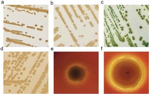
Table 4. Summary of physiological and biochemical characteristics of seven common species of the genus Shewanella.